. 2007 Aug;5(8):e209.
doi: 10.1371/journal.pbio.0050209.

Structure of the chloroplast ribosome: novel domains for translation regulation

Affiliations

Structure of the chloroplast ribosome: novel domains for translation regulation

Andrea L Manuell et al. PLoS Biol. 2007 Aug.

Abstract

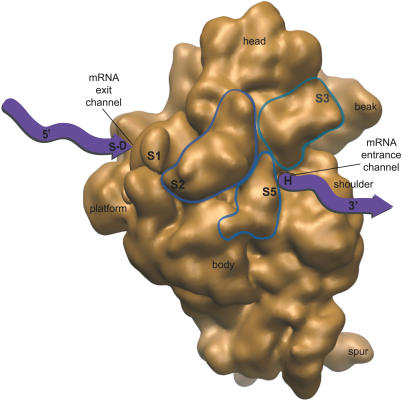
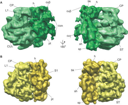
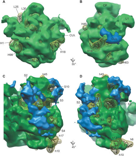
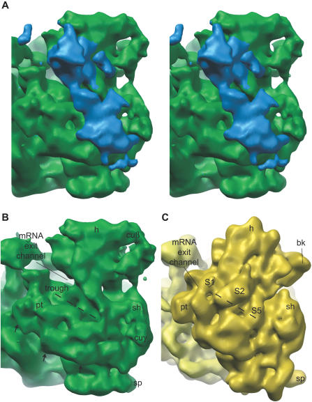
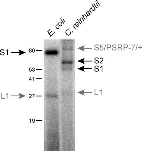
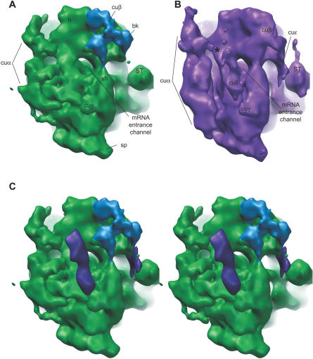
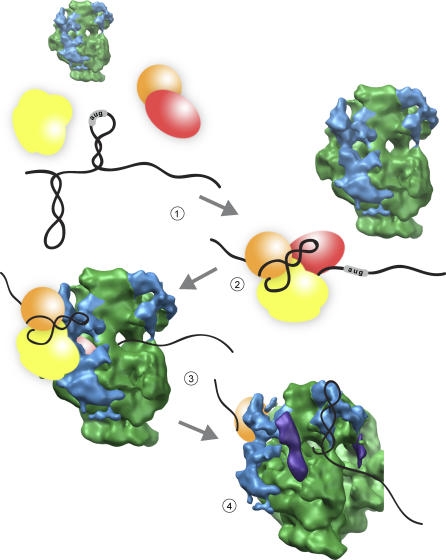
Gene expression in chloroplasts is controlled primarily through the regulation of translation. This regulation allows coordinate expression between the plastid and nuclear genomes, and is responsive to environmental conditions. Despite common ancestry with bacterial translation, chloroplast translation is more complex and involves positive regulatory mRNA elements and a host of requisite protein translation factors that do not have counterparts in bacteria. Previous proteomic analyses of the chloroplast ribosome identified a significant number of chloroplast-unique ribosomal proteins that expand upon a basic bacterial 70S-like composition. In this study, cryo-electron microscopy and single-particle reconstruction were used to calculate the structure of the chloroplast ribosome to a resolution of 15.5 A. Chloroplast-unique proteins are visualized as novel structural additions to a basic bacterial ribosome core. These structures are located at optimal positions on the chloroplast ribosome for interaction with mRNAs during translation initiation. Visualization of these chloroplast-unique structures on the ribosome, combined with mRNA cross-linking, allows us to propose a model for translation initiation in chloroplasts in which chloroplast-unique ribosomal proteins interact with plastid-specific translation factors and RNA elements to facilitate regulated translation of chloroplast mRNAs.


Conflict of interest statement

Competing interests. The authors have declared that no competing interests exist.

Figures

Figure 1
Figure 1. Predicted Location of Chloroplast-Unique Structures and Their Proximity to Functionally Important Regions of the Small Ribosomal Subunit
The solvent-exposed surface of a bacterial small subunit [31] is shown and faces the reader. Locations of small subunit ribosomal proteins that have large additional domains on the chloroplast ribosome (S2, S3, and S5) [26] are circled. These proteins are labeled at the predicted location of chloroplast-unique domains, based on sequence homology with bacterial orthologs and bacterial ribosome structure; the S3 label is on the back side of the circled protein, facing the beak. mRNA (purple ribbon) enters and exits the small subunit through discrete channels that are the sites of important functions of the ribosome (S-D, S-D interaction; H, helicase activity; see text). Chloroplast-unique domains from S2, S3, and S5 are predicted to form a structural feature on the chloroplast ribosome that is positioned in proximity to mRNA-interacting regions of the small subunit.
Figure 2
Figure 2. Three-Dimensional Map of the C. reinhardtii Chloroplast Ribosome at 15.5 Å
(A) Chloroplast ribosome (green). Compared to (B) bacterial ribosome cryoEM structure (yellow; [29]). Left and right views are related by 180° rotation as indicated. At left is a classic side view of the ribosome with the large subunit on the left and the small subunit on the right; right view shows the small subunit on the left and the large subunit on the right. Common ribosome landmarks as well as chloroplast-unique structures are clearly defined on the chloroplast ribosome, and have been labeled. bk, beak; CP, central protuberance; cuα, cuβ, cuγ, and CUλ, chloroplast-unique structures; h, head; L1, L1 arm; pt, platform; sh, shoulder; sp, spur; ST, stalk. See Video S1 for a three-dimensional view of the chloroplast ribosome structure.
Figure 3
Figure 3. Structural Differences between Chloroplast and Bacterial Ribosomes
Shown are differences between chloroplast and E. coli large ([A] and [B]) and small ([C] and [D]) subunits highlighting chloroplast-unique structures in blue, and E. coli ribosome densities lacking from the chloroplast ribosome in yellow mesh. Ribbon underlay of X-ray structure allows identification of missing densities. Landmarks as in Figure 2, plus some helix (e.g., H45) and protein (e.g., L25) labels. See text, Figures S3 and S4, and Tables S1 and S2 for specifics on these helices and proteins. (A) Solvent-exposed face of the large subunit. Helices and proteins that are not present on the chloroplast ribosome are clearly identified in difference density. (B) Rotated from (A) as indicated. The largest chloroplast-unique density on the large subunit is at the base of the L1 arm (CUλ). (C and D) Solvent-exposed face of the small subunit. See text for a discussion of each main chloroplast-unique structure (cuα and cuβ).
Figure 4
Figure 4. Chloroplast-Unique Structures Dominate the mRNA Exit Channel Area
Solvent-exposed face of the small subunit is shown slightly turned to look at the mRNA exit channel. (A) Stereo pair image. The largest chloroplast-unique structure on the ribosome, cuα (blue), emerges from the head and neck of the small subunit and partially defines the path of access for mRNA at the exit channel. (B) cuα has been removed from this image to reveal the trough from the mRNA exit channel down into the midbody of the small subunit (dashed line). The platform is slightly lifted on the chloroplast ribosome, as indicated by the black arrows (arrows not to scale), accentuating the trough. (C) E. coli ribosome cryoEM structure for comparison indicates the location of proteins known to be altered in the chloroplast ribosome along the path of the trough (S1, S2, and S5; see Figure S3).
Figure 5
Figure 5. Cross-Linking of Plastid mRNA 5′ UTR to Chloroplast and E. coli Ribosomes
Radiolabeled psbA 5′ UTR was incubated with purified chloroplast or E. coli ribosomes, UV irradiated to cross-link mRNA, and then proteins were separated by SDS-PAGE. Protein size markers are indicated (in kDa). In E. coli, S1 is the main protein that is cross-linked to mRNA, while a small amount is cross-linked to another protein, presumably L1 (gray arrow on left lane). Chloroplast ribosomes have two proteins that clearly bind to mRNA, S1 and S2; two other bands are also labeled (gray arrows on right lane). One of these bands is L1, and the other appears as an incompletely denatured protein complex containing at least S5 and PSRP-7. Mass spectrometry was used to identify the protein component of labeled chloroplast ribosome bands (see Materials and Methods).
Figure 6
Figure 6. Chloroplast-Unique Density Approaches the mRNA Entrance Channel
Solvent-exposed face of the small subunit is shown slightly turned to look at the mRNA entrance channel. (A) cuβ (blue) connects the beak helix with the head, squaring out this normally pointed feature on the ribosome, and approaches the top of the mRNA entrance channel. Connection to the shoulder stems from this chloroplast-unique structure as well. (B) PSRP-7 antibody-labeled chloroplast ribosome structure has additional density near the mRNA entrance channel (cuδ) and extending from the beak helix toward the factor-binding site (cuɛ). (C) Stereo pair image (putative PSRP-7 density labeled in purple) shows the position of cuδ across the mRNA entrance channel and paralleling cuα off the solvent-exposed face of the small subunit.
Figure 7
Figure 7. Translation Initiation in the Chloroplast
Chloroplast mRNAs are not competent for translation initiation until assembled with mRNA-specific translation factors (panel 1). Translation factors assembled on the 5′ UTR of a chloroplast message modify the secondary structure of the mRNA and form a novel translation initiation mRNP (panel 2). This mRNP complex binds to the chloroplast ribosome through interaction with the chloroplast-unique structure (cuα, shown in blue) around the mRNA exit channel (panel 3). This binding positions the start site AUG of the mRNA in the P-site for translation initiation. During elongation, signal elements in the mRNA may be recognized by chloroplast-unique structures around the mRNA entrance channel like cuβ (panel 4). Such binding may modify the processivity of translation of that mRNA, and facilitate pausing to allow for cotranslational membrane insertion or cofactor assembly.

References

    1. Sugiura M. The chloroplast genome. Plant Mol Biol. 1992;19:149–168. - PubMed
    1. Watson JC, Surzycki SJ. Both the chloroplast and nuclear genome of Chlamydomonas reinhardtii share homology with Escherichia coli genes for transcriptional and translation components. Curr Genet. 1983;7:201–210. - PubMed
    1. Daniell H, Chebolu S, Kumar S, Singleton M, Falconer R. Chloroplast-derived vaccine antigens and other therapeutic proteins. Vaccine. 2005;23:1779–1783. - PubMed
    1. Maul JE, Lilly JW, Cui L, dePamphilis CW, Miller W, et al. The Chlamydomonas reinhardtii plastid chromosome: Islands of genes in a sea of repeats. Plant Cell. 2002;14:2659–2679. - PMC - PubMed
    1. Barkan A, Goldschmidt-Clermont M. Participation of nuclear genes in chloroplast gene expression. Biochimie. 2000;82:559–572. - PubMed

Publication types

MeSH terms