. 2007 Oct;98(10):1609-16.
doi: 10.1111/j.1349-7006.2007.00571.x. Epub 2007 Aug 7.

Pilot study of MDR1 gene transfer into hematopoietic stem cells and chemoprotection in metastatic breast cancer patients

Affiliations

Pilot study of MDR1 gene transfer into hematopoietic stem cells and chemoprotection in metastatic breast cancer patients

Shunji Takahashi et al. Cancer Sci. 2007 Oct.

Abstract

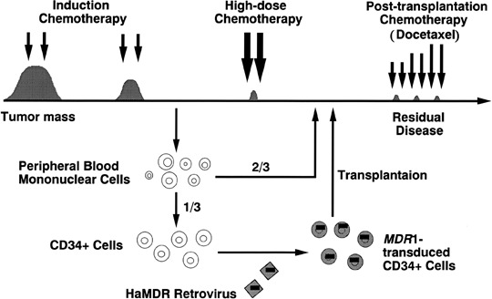
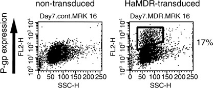
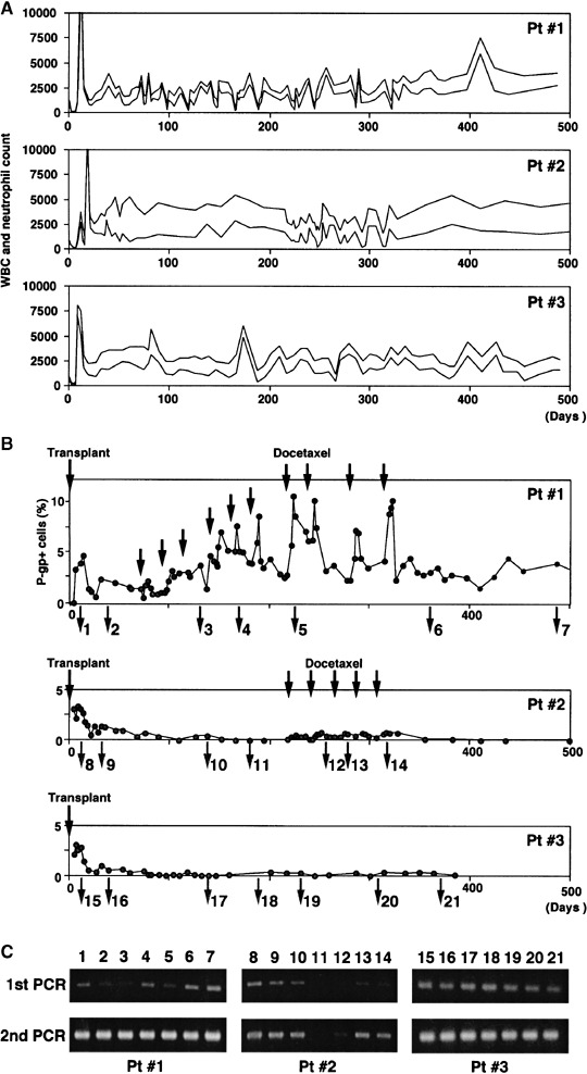
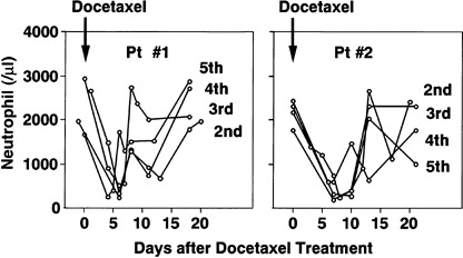
A major problem in high-dose chemotherapy with autologous hematopoietic stem cell transplantation is insufficient function of reconstituted bone marrow that limits the efficacy of post-transplantation chemotherapy. Because transduction of hematopoietic stem cells with the multidrug resistance 1 (MDR1) gene might circumvent this problem, we conducted a pilot study of MDR1 gene therapy against metastatic breast cancer. Peripheral blood stem cells were harvested, and one-third of the cells were transduced with MDR1 retrovirus. After the reconstitution of bone marrow function, the patients received high-dose chemotherapy with transplantation of both MDR1-transduced and unprocessed peripheral blood stem cells. The patients then received docetaxel chemotherapy. Two patients received transplantation of the MDR1-transduced cells in 2001. Peripheral blood MDR1-transduced leukocytes were 3-5% of the total cells after transplantation, but decreased gradually. During docetaxel chemotherapy, an increase in the rate of MDR1-transduced leukocytes (up to 10%) was observed. Comparison of docetaxel-induced granulocytopenia in the two patients suggested a bone marrow-protective effect of the MDR1-transduced cells. No serious side-effect was observed, and the patients were in complete remission for more than 3 years. The MDR1-transduced cells gradually decreased and disappeared almost entirely by the end of 2004. Results of linear amplification-mediated polymerase chain reaction of the MDR1-transduced leukocytes suggested no sign of abnormal amplification of the transduced cells. A third patient received transplantation of the MDR1-transduced cells in 2004. These results suggest the feasibility of our MDR1 gene therapy against metastatic breast cancer, and follow-up is ongoing.


Figures

Figure 1
Figure 1
The outline of multidrug resistance 1 (MDR1) gene therapy. After remission induction chemotherapy for metastatic breast cancer, peripheral blood stem cells (PBSC) were harvested from patients in remission, and one‐third were transduced with retroviral MDR1 vector. The patients then received high‐dose chemotherapy with transplantation of both MDR1‐transduced and unprocessed PBSC. After bone marrow reconstitution, the patients received docetaxel chemotherapy.
Figure 2
Figure 2
Ex vivo multidrug resistance 1 (MDR1) transduction of peripheral blood stem cells (PBSC) in patient 1. CD34‐positive cells were isolated from freshly harvested PBSC of the patients using an Isolex 50 Stem Cell Reagent kit. The CD34‐enriched cells were then stimulated with granulocyte colony‐stimulating factor (G‐CSF), thrombopoietin (TPO), interleukin (IL)‐6, sIL‐6R and Flk2/Flt3‐ligand (FL‐ligand), and transduced with HaMDR viral supernatant. Non‐transduced or HaMDR‐transduced cells were incubated with the F(ab′)2 fragment of MRK16, and were washed and incubated with R‐phycoerythrin‐conjugated streptavidin. The fluorescence staining level was analyzed using FACScan. The analysis was carried out after 2 days of stem cell amplification, 2 days of viral transfer, and then 2 days of further culture. P‐gp, P‐glycoprotein.
Figure 3
Figure 3
Changes in (A) white blood cells (WBC) and neutrophils, and (B) P‐glycoprotein (P‐gp)‐positive cells after peripheral blood stem cell transplantation (PBSCT) and (C) semiquantitative polymerase chain reaction (PCR) of HaMDR DNA in peripheral blood cells of the three patients. Docetaxel chemotherapy was started 60 days after PBSCT in patient 1 and 208 days after PBSCT in patient 2. The patients were then treated with 30 mg/m2 (first course) or 45 mg/m2 (second course and later) docetaxel (black arrow). (A) Plots of total white blood cells (upper line) and neutrophils (lower line). (B) Mononuclear cells (MNC) were separated from peripheral blood by Ficoll‐Hypaque density gradient centrifugation, incubated with the F(ab′)2 fragment of MRK16, and washed and incubated with R‐phycoerythrin‐conjugated streptavidin. The fluorescence staining level was analyzed using FACScan. (C) Genomic DNA was extracted from peripheral blood of the three patients at various time points. Multidrug resistance 1 (MDR1) genes were amplified with the PCR primers for 40 cycles. Then the second PCR was carried out with the same primers. PCR products were analyzed on agarose gels. Patient 1: 1, day 12 after PBSCT; 2, day 40; 3, day 131 (after third docetaxel chemotherapy); 4, day 170 (after fifth docetaxel); 5, day 226 (after seventh docetaxel); 6, day 362 (after 10th docetaxel); 7, day 488. Patient 2: 8, day 12 after PBSCT; 9, day 33; 10, day 138; 11, day 170; 12, day 247 (after second docetaxel chemotherapy); 13, day 268 (after third docetaxel); 14, day 310 (after fifth docetaxel). Patient 3: 15, day 12 after PBSCT; 16, day 40; 17, day 139; 18, day 188; 19, day 231; 20, day 308; 21, day 371.
Figure 4
Figure 4
Comparison of neutrophil count after docetaxel chemotherapy in patients 1 and 2. The changes in neutrophil count after the second to fifth docetaxel chemotherapy (45 mg/m2) in patients 1 and 2 are shown.
Figure 5
Figure 5
Linear amplification‐mediated (LAM)‐polymerase chain reaction (PCR) analysis of HaMDR integration sites in the peripheral blood leukocytes of patient 1. Genomic DNA was extracted from peripheral blood of the first patient at various time points. The genomic–proviral junction sequence was preamplified by repeated primer. Biotinylated extension products were selected, treated with Klenow fragment and incubated with MaeII. The product was then treated with T4 DNA Ligase and amplified with AmpliTaq Gold DNA polymerase with vector‐specific primer and linker cassette primer, then further amplified with the same primer set. PCR products were analyzed on agarose gels. 1, day 12 after PBSCT; 2, day 40; 3, day 112 (after second docetaxel chemotherapy); 4, day 152 (after fourth docetaxel); 5, day 196 (after sixth docetaxel); 6, day 257 (after eighth docetaxel); 7, day 327 (after 10th docetaxel); 8, day 586; 9, day 727; 10, day 762; 11, day 790.

Similar articles

Cited by

References

    1. Gregory WM, Smith P, Richards MA, Twelves CJ, Knight RK, Rubens RD. Chemotherapy of advanced breast cancer: outcome and prognostic factors. Br J Cancer 1993; 68: 988–95. - PMC - PubMed
    1. Inoue K, Ogawa M, Horikoshi N et al . Evaluation of prognostic factors for 233 patients with recurrent advanced breast cancer. Jpn J Clin Oncol 1991; 21: 334–9. - PubMed
    1. Philip T, Guglielmi C, Hagenbeek A et al . Autologous bone marrow transplantation as compared with salvage chemotherapy in relapses of chemotherapy‐sensitive non‐Hodgkin's lymphoma. N Engl J Med 1995; 333: 1540–5. - PubMed
    1. Attal M, Harousseau JL, Stoppa AM et al . A prospective, randomized trial of autologous bone marrow transplantation and chemotherapy in multiple myeloma. Intergroupe Francais du Myelome. N Engl J Med 1996; 335: 91–7. - PubMed
    1. Ito Y, Mukaiyama T, Ogawa M et al . Epirubicin‐containing high‐dose chemotherapy followed by autologous hematopoietic progenitor cell transfusion for patients with chemotherapy‐sensitive metastatic breast cancer: results of 5‐year follow‐up. Cancer Chemother Pharmacol 1999; 43: 8–12. - PubMed

Publication types

MeSH terms

Substances