Case report: PET/CT, a cautionary tale
- PMID: 17683560
- PMCID: PMC1950887
- DOI: 10.1186/1471-2407-7-147
Case report: PET/CT, a cautionary tale
Abstract
Background: The use of combined positron emission tomography/computerised tomography (PET/CT) scanners in oncology has been shown to improve the staging of tumours and the detection of relapses. However, mis-registration errors are increasingly recognised to be a common pitfall of PET/CT studies.
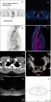
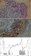
Case presentation: We report a patient with a germ cell tumour of the testis, who underwent a PET/CT scan to detect the site of relapse with a view to surgical removal. However, the PET/CT scan mislocalised the tumour site to be within the T2 vertebral body. A subsequent endoscopic ultrasound scan however showed the tumour to be anterior to the vertebral body, which was confirmed at surgery.
Conclusion: In this report, we highlight the artefactual mislocalisation errors which may occur with PET/CT imaging, and the need to review and verify these scans.
Figures


Similar articles
-
Early prediction of response to chemoradiotherapy for head and neck cancer: reliability of restaging with combined positron emission tomography and computed tomography.Arch Otolaryngol Head Neck Surg. 2009 Nov;135(11):1119-25. doi: 10.1001/archoto.2009.152. Arch Otolaryngol Head Neck Surg. 2009. PMID: 19917925
-
Imaging of testicular germ cell tumours.Cancer Imaging. 2006 Sep 7;6(1):124-34. doi: 10.1102/1470-7330.2006.0020. Cancer Imaging. 2006. PMID: 16966068 Free PMC article.
-
PET/CT for the staging and follow-up of patients with malignancies.Eur J Radiol. 2009 Jun;70(3):382-92. doi: 10.1016/j.ejrad.2009.03.051. Epub 2009 Apr 29. Eur J Radiol. 2009. PMID: 19406595 Review.
-
Whole-body magnetic resonance imaging and positron emission tomography-computed tomography in oncology.Top Magn Reson Imaging. 2007 Jun;18(3):193-202. doi: 10.1097/RMR.0b013e318093e6bo. Top Magn Reson Imaging. 2007. PMID: 17762383 Review.
-
Is positron emission tomography useful in locoregional staging of esophageal cancer? Results of a multidisciplinary initiative comparing CT, positron emission tomography, and EUS.Gastrointest Endosc. 2008 Mar;67(3):402-9. doi: 10.1016/j.gie.2007.09.006. Gastrointest Endosc. 2008. PMID: 18178202
References
-
- Som P, Atkins HL, Bandoypadhyay D, Fowler JS, MacGregor RR, Matsui K, Oster ZH, Sacker DF, Shiue CY, Turner H, Wan CN, Wolf AP, Zabinski SV. A fluorinated glucose analog, 2-fluoro-2-deoxy-D-glucose (F-18): nontoxic tracer for rapid tumor detection. J Nucl Med. 1980;21:670–675. - PubMed
-
- Beyer T, Townsend DW, Brun T, Kinahan PE, Charron M, Roddy R, Jerin J, Young J, Byars L, Nutt R. A combined PET/CT scanner for clinical oncology. J Nucl Med. 2000;41:1369–1379. - PubMed
-
- Metser U, Golan O, Levine CD, Even-Sapir E. Tumor lesion detection: when is integrated positron emission tomography/computed tomography more accurate than side-by-side interpretation of positron emission tomography and computed tomography? J Comput Assist Tomogr. 2005;29:554–559. doi: 10.1097/01.rct.0000164671.96143.c2. - DOI - PubMed

