. 2007 Aug 6:8:33.
doi: 10.1186/1471-2121-8-33.

Establishment and mitotic stability of an extra-chromosomal mammalian replicon

Affiliations

Establishment and mitotic stability of an extra-chromosomal mammalian replicon

Isa M Stehle et al. BMC Cell Biol. .

Abstract

Background: Basic functions of the eukaryotic nucleus, like transcription and replication, are regulated in a hierarchic fashion. It is assumed that epigenetic factors influence the efficiency and precision of these processes. In order to uncouple local and long-range epigenetic features we used an extra-chromosomal replicon to study the requirements for replication and segregation and compared its behavior to that of its integrated counterpart.

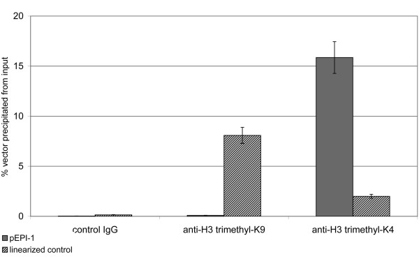
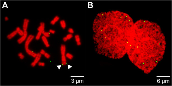
Results: The autonomous replicon replicates in all eukaryotic cells and is stably maintained in the absence of selection but, as other extra-chromosomal replicons, its establishment is very inefficient. We now show that following establishment the vector is stably associated with nuclear compartments involved in gene expression and chromosomal domains that replicate at the onset of S-phase. While the vector stays autonomous, its association with these compartments ensures the efficiency of replication and mitotic segregation in proliferating cells.

Conclusion: Using this novel minimal model system we demonstrate that relevant functions of the eukaryotic nucleus are strongly influenced by higher nuclear architecture. Furthermore our findings have relevance for the rational design of episomal vectors to be used for genetic modification of cells: in order to improve such constructs with respect to efficiency elements have to be identified which ensure that such constructs reach regions of the nucleus favorable for replication and transcription.


Figures

Figure 1
Figure 1
(A) Map of pEPI-EGFP. pEPI-EGFP derives from the commercial plasmid pGFP-C1 (Clontech). An S/MAR sequence, obtained from the human IFN β-gene, was cloned into the multiple cloning site (MCS) of pGFP-C1 [7] resulting in the vector pEPI-1. The GFP gene was substituted by the enhanced version EGFP in pEPI-EGFP. The Neo/Kan gene is driven by dual promoters to confer kanamycin resistance in bacteria and G418 resistance in mammalian cells. BglII and EcoRI restriction sites are indicated. SV40, simian virus 40; ori, origin of replication; HSV, herpes simplex virus. (B) Southern blot analysis of extra-chromosomal DNA and integrated vector copies using pEPI as a probe. Lane1: 1-Kbp Ladder (O'Gene Ruler; Fermentas, St. Leon-Rot, Germany). Lane2: free extra-chromosomal pEPI DNA isolated from CHO cells transfected with supercoiled plasmid DNA, linearized by digestion with BglII demonstrating the episomal state of the vector. Lane3 and 4: DNA isolated from CHO cells transfected with linearized plasmid DNA. In this case the vector integrates at random sites into the genome and can become rearranged. DNA was digested with BglII.
Figure 2
Figure 2
Establishment of an autonomous replicon. Transfection of CHO cells with pEPI or linearized vectors was performed using the lipid-based transfection reagent FuGENE 6 (Roche). Subsequently the establishment of the episome in the cell nucleus was monitored 6 h (A), 24 h (B) and after a selection period of 10 days (C, D) post transfection. The episome (green) was visualized by pEPI FISH. To-Pro-3 was used for DNA counterstaining (red) in (A-D). Maximum intensity projections were rendered from a set of 5 mid serial sections. The cellular contour was outlined with a white line in (A, B). (A) Vesicles containing numerous vector molecules occurred within the cytoplasm 6 h post transfection, whereas no signals were observed within the nucleus. (B) A strong intranuclear pEPI signal indicating a large number of vector molecules was observed 12 h post transfection, while cytoplasmic localization of pEPI was no longer observed. (C) In the majority of cases, cells transfected with the linearized vectors displayed FISH signals consistent with integration of the vector at a single chromosomal locus (D) Solely a limited number of distinct pEPI signals was observed after a selection period of 10 days suggesting that only a minor portion of the episome is stably established.
Figure 3
Figure 3
Localization of the episome in interphase nuclei. The qualitative co-localization of the episome with subnuclear structures in interphase nuclei was analyzed using pEPI FISH (A-J). In some experiments pEPI FISH was used in combination with immunofluorescence techniques to analyse the co-localization of pEPI with subnuclear marker proteins (E-J). Co-localizing voxels of two channels being analyzed were highlighted in white color in (A, B, C, D, H). The episome (green) was visualized by pEPI FISH. To-Pro-3 was used for DNA counterstaining (red) in (A, B, C, D, E, G, I). Maximum intensity projections were rendered from a set of 5 mid serial sections (A, B, C, D, E, G, H, I). For further details see Additional file 2. (A, B) Co-localization analyses showed that cells transfected with the linearized vectors displayed FISH signals consistent with integration of the vector at a single chromosomal locus as the vectors completely co-localized with domains containing condensed chromatin. Equal results were obtained when hyper-condensation of chromatin was induced prior to fixation (B). (C, D) Analyses of single pEPI signals revealed that the vector did not (green) or at most incompletely (white/green) colocalize to condensed chromatin regions (red). Insets are 400% magnifications of white or blue framed sectors in C and D. Such representative pEPI signals consist of green voxels (negative co-localization with condensed chromatin) or are composed of white and green voxels (incomplete co-localization with condensed chromatin). Negative or incomplete co-localization suggests that the episome is localized within the IC or at perichromatin regions bordering the IC at condensed chromatin surfaces. Equal results were obtained when hyper-condensation of chromatin was induced prior to fixation (B). (For selected light optical sections corresponding to the nuclei displayed in C and D as maximum intensity projections see the Additional file 2.) (E, F) Co-localization analyses of the episome (green) and SC35 (blue) showed that the episome occured co-localized or in close proximity to nuclear speckles, a structure which is found within the IC. (F) A 3D reconstruction was rendered from the same nucleus as displayed in (E). (G, H) Nuclear localization of the episome (green) and histone3 acetylated at lysines 9/14 (H3 acetyl-K9/K14) (blue). The nuclear counterstain (red) channel was hidden in (H) to facilitate co-localization analysis of the episome and H3 acetyl-K9/K14 showing that the episome occurred co-localized or in close proximity to sites of active transcription. (I, J) Co-localization analyses of the episome (green) and histone3 trimethylated at lysine9 (H3 trimethyl-K9) (blue) showed that the episome did not co-localize with such sites. (J) A 3D reconstruction was rendered from the same nucleus as displayed in (I). Bar in (A) is representative for (A-J).
Figure 4
Figure 4
Quantitative real time PCR analyses of ChIP experiments. ChIP experiments were performed using antibodies targeted to H3 trimethyl-K9 (condensed/inactive chromatin: mid columns), H3 trimethyl-K4 (open/active chromatin: right handed columns) or using a control IgG (left handed columns). The percentage of either pEPI-1 (gray columns) or the linearized control vector (striped columns) precipitated from input was quantified using real time PCR. Since no significant amount of the vector molecules was pulled down with the control IgG, it could be clearly demonstrated that about 30 times more vector molecules were precipitated using the anti-H3 trimethyl-K9 antibody in a precipitate from cells containing the integrated vector compared to cells containing the vector in its episomal form. When the same chromatin preparations were precipitated with an antibody directed against H3 trimethyl-K4 over 20 times more vector molecules were obtained from cells containing the episome compared to cells containing the integrated vector.
Figure 5
Figure 5
Association of the episome with metaphase chromosomes. The localization of the episome was studied by FISH on spreads of metaphase chromosomes (A), and the equal distribution of vector molecules was monitored in postmitotic nuclei of dividing cells (B). The episome (green) was visualized by pEPI FISH. To-Pro-3 was used for DNA counterstaining (red). A maximum intensity projection was rendered from a set of 5 mid serial sections in (B). Arrows in (A) indicate a pEPI signal pair, each signal localized on a sister chromatid. For further details see Additional file 4.
Figure 6
Figure 6
Co-localization of the episome with early replication foci. The qualitative co-localization of the episome and replication foci was analyzed using pEPI FISH and BrdU pulse labeling. Maximum intensity projections were rendered from a set of serial sections. The nuclear contour was outlined with a white line. (A) Replication foci were labeled during early S-phase by incorporation of BrdU (1 h) into newly synthesized DNA. Subsequently nuclei were fixed and BrdU (red) was visualized by antibody staining. The episome (green) was visualized by pEPI FISH. Co-localizing voxels were highlighted in white color. (B) Alternatively nascent DNA was pulse-labeled with BrdU for 1 h during early S-phase, fixing and subsequent immunolocalization was done following cell division. The episome (green) was visualized by pEPI FISH. Co-localizing voxels were highlighted in white color.

Similar articles

Cited by

References

    1. Cremer T, Cremer M, Dietzel S, Muller S, Solovei I, Fakan S. Chromosome territories - a functional nuclear landscape. Curr Opin Cell Biol. 2006;18:307–316. doi: 10.1016/j.ceb.2006.04.007. - DOI - PubMed
    1. Khorasanizadeh S. The nucleosome: from genomic organization to genomic regulation. Cell. 2004;116:259–272. doi: 10.1016/S0092-8674(04)00044-3. - DOI - PubMed
    1. Akhtar A, Matera AG. In and around the nucleus. Nat Cell Biol. 2006;8:3–6. doi: 10.1038/ncb0106-3. - DOI - PubMed
    1. Cook PR. The organization of replication and transcription. Science. 1999;284:1790–1795. doi: 10.1126/science.284.5421.1790. - DOI - PubMed
    1. Baiker A, Maercker C, Piechaczek C, Schmidt SB, Bode J, Benham C, Lipps HJ. Mitotic stability of an episomal vector containing a human scaffold/matrix-attached region is provided by association with nuclear matrix. Nat Cell Biol. 2000;2:182–184. doi: 10.1038/35004061. - DOI - PubMed

Publication types

LinkOut - more resources