. 2007 Aug 6:7:94.
doi: 10.1186/1471-213X-7-94.

Analysis of the Rana catesbeiana tadpole tail fin proteome and phosphoproteome during T3-induced apoptosis: identification of a novel type I keratin

Affiliations

Analysis of the Rana catesbeiana tadpole tail fin proteome and phosphoproteome during T3-induced apoptosis: identification of a novel type I keratin

Dominik Domanski et al. BMC Dev Biol. .

Abstract

Background: Thyroid hormones (THs) are vital in the maintenance of homeostasis and in the control of development. One postembryonic developmental process that is principally regulated by THs is amphibian metamorphosis. This process has been intensively studied at the genomic level yet very little information at the proteomic level exists. In addition, there is increasing evidence that changes in the phosphoproteome influence TH action.

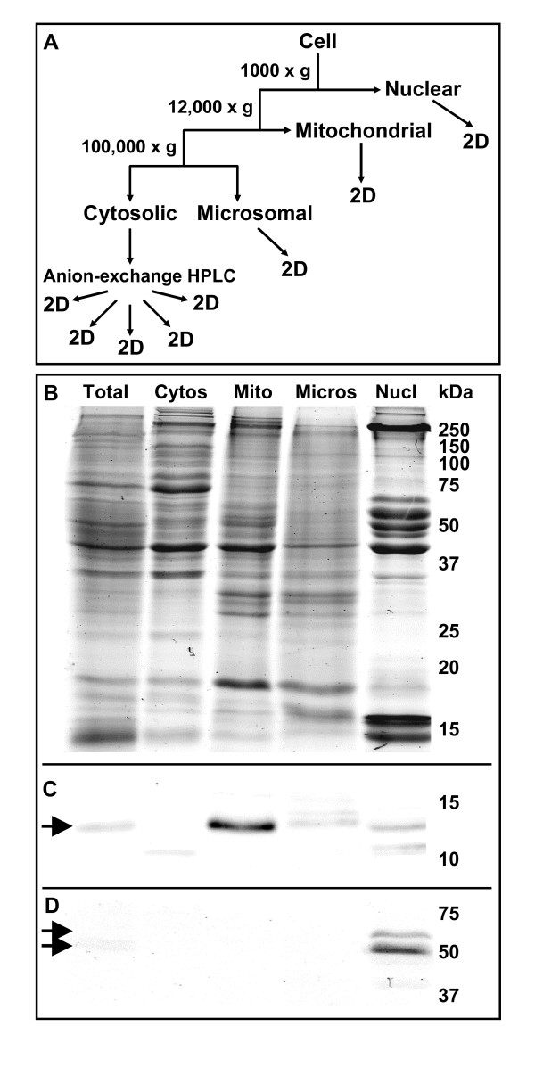
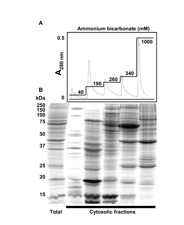
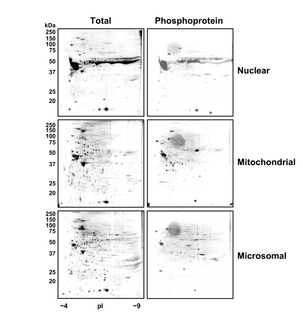
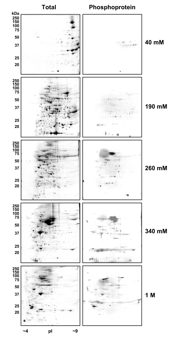
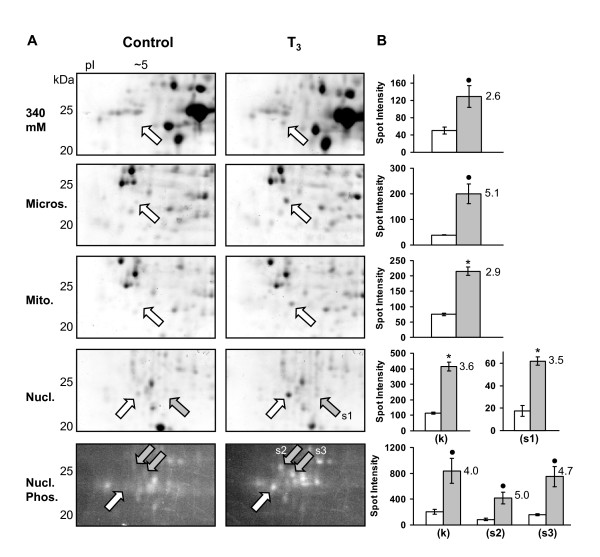
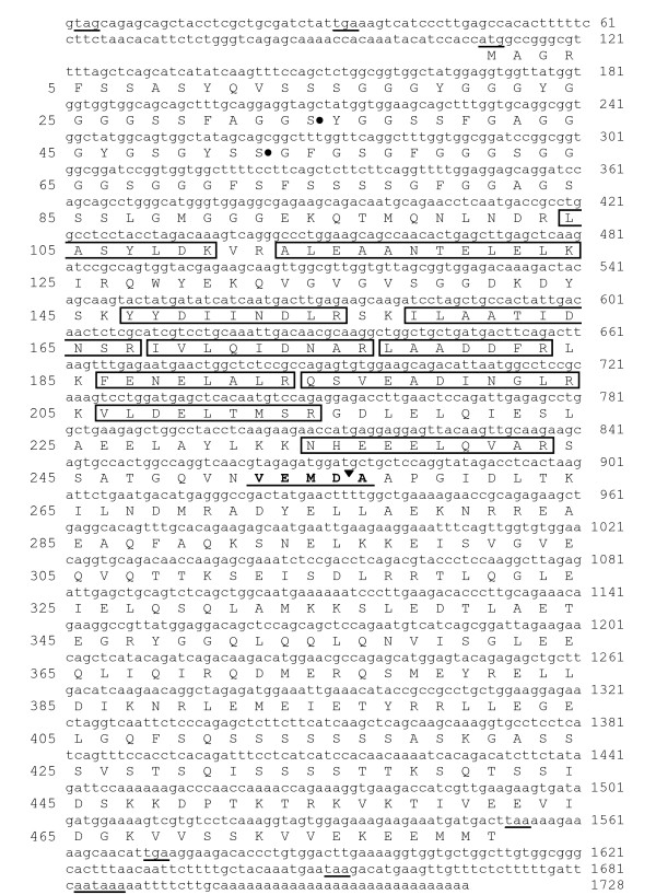
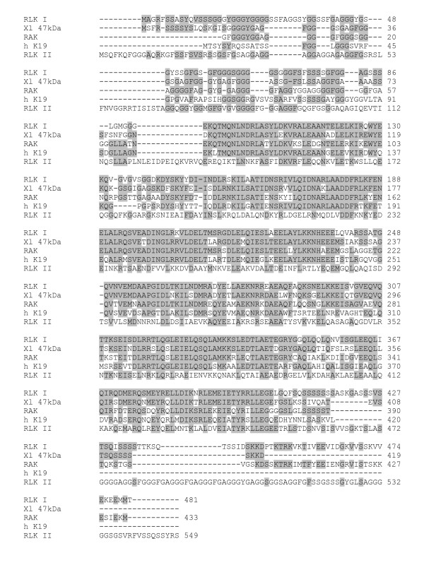
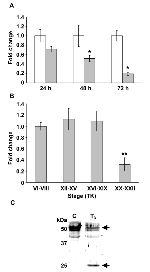
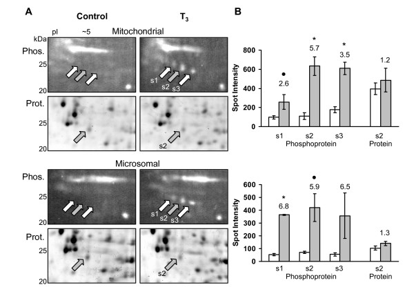
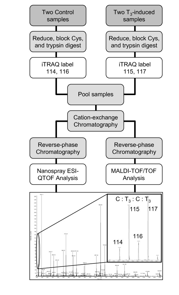
Results: Here we identify components of the proteome and phosphoproteome in the tail fin that changed within 48 h of exposure of premetamorphic Rana catesbeiana tadpoles to 10 nM 3,5,3'-triiodothyronine (T3). To this end, we developed a cell and protein fractionation method combined with two-dimensional gel electrophoresis and phosphoprotein-specific staining. Altered proteins were identified using mass spectrometry (MS). We identified and cloned a novel Rana larval type I keratin, RLK I, which may be a target for caspase-mediated proteolysis upon exposure to T3. In addition, the RLK I transcript is reduced during T3-induced and natural metamorphosis which is consistent with a larval keratin. Furthermore, GILT, a protein involved in the immune system, is changed in phosphorylation state which is linked to its activation. Using a complementary MS technique for the analysis of differentially-expressed proteins, isobaric tags for relative and absolute quantitation (iTRAQ) revealed 15 additional proteins whose levels were altered upon T3 treatment. The success of identifying proteins whose levels changed upon T3 treatment with iTRAQ was enhanced through de novo sequencing of MS data and homology database searching. These proteins are involved in apoptosis, extracellular matrix structure, immune system, metabolism, mechanical function, and oxygen transport.

Conclusion: We have demonstrated the ability to derive proteomics-based information from a model species for postembryonic development for which no genome information is currently available. The present study identifies proteins whose levels and/or phosphorylation states are altered within 48 h of the induction of tadpole tail regression prior to overt remodeling of the tail. In particular, we have identified a novel keratin that is a target for T3-mediated changes in the tail that can serve as an indicator of early response to this hormone.


Figures

Figure 1
Figure 1
Subcellular fractionation of the tail fin proteome. (A) Fractionation of tail fin cells into subcellular compartments and subsequent treatments of those fractions. Two different extraction procedures, based on differential centrifugation, were developed to generate the nuclear and the cytoplasmic/mitochondrial/microsomal fractions. (B) SDS-PAGE shows the successful fractionation of the total tail fin proteome into the cytosolic (Cytos), mitochondrial (Mito), microsomal (Micros), and nuclear (Nucl) fractions. Relative molecular weights of protein standards are indicated in kDa. (C) Immunoblot of the gel in (B) for the mitochondrial marker, cytochrome c (arrow) showing the enrichment of mitochondria in the expected fraction. (D) Immunoblot of the gel in (B) for the nuclear markers, lamin B1 and B2 (double arrow) showing the enrichment of nuclei in the expected fraction.
Figure 2
Figure 2
Anion-exchange HPLC fractionation of the cytosolic fraction. (A) The cytosolic fraction was further fractionated using an anion-exchange column (Accell QMA) with a step-gradient of increasing concentrations of ammonium bicarbonate (straight lines). The concentrations are indicated on each step while absorbance was measured at 280 nm indicating the protein yield of each fraction. (B) The Coomassie blue-stained SDS-PAGE gel shows the fractionation of the cytosolic sample (total) with the lanes corresponding to the cytosolic fractions below the peaks of the HPLC chromatogram. Note the resulting enrichment of certain protein bands. Relative molecular weights of protein standards are indicated in kDa.
Figure 3
Figure 3
2D gel analyses of the nuclear, mitochondrial and microsomal fractions. Proteins from the nuclear, mitochondrial and microsomal fractions were separated by 2D-PAGE according to molecular weight and pI point. Total proteins were detected by colloidal Coomassie stain while phosphoproteins were detected in the same gel using the ProQ Diamond phosphoprotein-specific stain. Relative molecular weights of protein standards are indicated in kDa.
Figure 4
Figure 4
2D gel analysis of the anion-exchange HPLC cytosolic fractions. Proteins from each of the fractions resulting from the anion-exchange HPLC of the cytosolic sample were separated by 2D-PAGE according to molecular weight and pI point. The 40 mM fraction is the unbound protein fraction, while the subsequent fractions are proteins eluted by the increasing ammonium bicarbonate concentration step-gradients. Total proteins were detected by colloidal Coomassie stain while phosphoproteins were detected in the same gel using the ProQ Diamond phosphoprotein-specific stain. Relative molecular weights of protein standards are indicated in kDa.
Figure 5
Figure 5
Identification of a novel R. catesbeiana type I (RLK I) keratin fragment by 2D gel analysis. (A) 2D gel regions of the 340 mM cytosolic, microsomal, mitochondrial, and nuclear fractions show the increase of a protein spot at ~24 kDa and pI ~5 due to T3 treatment at 48 h. The corresponding gel region, stained with a phosphoprotein stain, is shown for the nuclear fraction revealing additional changes in the phosphoproteome. The white arrows indicate the spot identified as a novel type I keratin RLK I fragment in the T3 samples (see Table 1). In the phosphoprotein gel, the white arrow indicates a possible phosphorylated form of the keratin fragment. The gray arrows indicate an additional unidentified protein and phosphoproteins that are altered upon T3 treatment. Relative molecular weights of protein standards are indicated in kDa. (B) Spot density measurements (in arbitrary values) are graphed for the corresponding 2D gels on the left. The white bar represents the control while the gray bar represents the T3 treatment. Error bars represent the standard error of the mean from three independent controls and three independent T3 samples. Significance is indicated by an asterisk for p < 0.01 and by a black dot for p < 0.04 (ANOVA). The values adjacent to the gray bars represent the fold increase due to T3. In the nuclear fraction (k) represents the keratin spot, while (s1) represents an additional protein spot observed to be increased, and (s2) and (s3) represent two phosphoproteins that were increased due to T3 treatment. Spot density measurements were normalized between the gels with the β-actin protein spot.
Figure 6
Figure 6
RLK I cDNA and derived amino acid sequence and location of MS/MS peptide fragments. The complete nucleotide sequence (lower case) of RLK I cDNA is shown. Underlined nucleotide sequences indicate all the in-frame stop codons, the first methionine codon, and a consensus AATAAA polyadenylation signal. Numbers on the right indicate nucleotide position. Upper case letters indicate the deduced amino acid sequence (single letter code). Boxed sequences indicate tryptic peptides observed in the MS analyses of the RLK I protein spot from the 2D analysis. The underlined VEMDA sequence indicates a consensus caspase cleavage site identified in human type I keratins with the black inverted-triangle indicating the cleavage site. Black dots adjacent to two serine residues indicate possible phosphorylation sites based on those found in human K18 at Ser33 and Ser52 (here Ser34 and Ser52). Numbers on the left indicate amino acid position.
Figure 7
Figure 7
Multiple sequence alignment of the derived amino acid sequence of RLK I. The derived amino acid sequence of RLK I [GenBank: EF156435] was aligned with the X. laevis type I keratin 47 kDa protein [GenBank: P05781] (Xl 47 kDa), a partial R. catesbeiana adult type I keratin RAK [GenBank: BAB47394.1] (RAK), the human type I keratin K19 [GenBank: NP_002267.2] (h K19), and the R. catesbeiana larval type II keratin RLK [GenBank: BAB47395] (RLK II) sequences. Gaps that were inserted for optimal alignment are indicated by a dash. Identical amino acids are shaded. Numbers indicate amino acid position for each sequence. The alignment was done using ClustalW software [22].
Figure 8
Figure 8
Changes in transcript and protein fragment levels of RLK I in the tail fin. (A) Fold change in steady-state levels of the keratin transcript relative to time-matched controls after 100 nM T3 exposure for 24, 48 and 72 h. White bars represent controls and gray bars represent T3 treatments. Error bars represent the standard error of the mean (n = 4 for all treatments). Significance is indicated by an asterisk for p < 0.03 (Mann-Whitney U). (B) Fold change in steady-state levels of the keratin transcript at different stages of natural metamorphosis relative to premetamorphic TK stage VI-VIII [31]. Error bars represent the standard error of the mean (n = 4 for all treatments). Significance is indicated by a double asterisk for p < 0.002 (Mann-Whitney U). (C) Immunobloting microsomal fraction samples using anti-pan-cytokeratin antibody reveals the appearance of the keratin fragment (25 kDa) and a concomitant loss of keratin at 50 kDa due to 10 nM T3 treatment. Relative molecular weights of protein standards are indicated in kDa. Shown is a representative of two independent experiments.
Figure 9
Figure 9
Phosphorylation changes in γ-interferon-inducible lysosomal thiol reductase (GILT). (A) Phosphoprotein 2D gel regions of the mitochondrial and microsomal fractions showing the increase in a row of phosphoprotein spots (s1, s2, s3) due to T3 treatment at 48 h, while a corresponding total-protein stained 2D region shows no change in the only detectable protein spot s2 (gray arrows). Relative molecular weights of protein standards are indicated in kDa. (B) Spot density measurements (in arbitrary values) are graphed for the corresponding 2D gels on the left. The white bar represents the control while the gray bar represents the T3 treatment. Error bars represent the standard error of the mean from three independent controls and three independent T3 samples. Significance is indicated by an asterisk for p < 0.01 and by black dot for p < 0.1 (ANOVA). The values adjacent to the gray bars represent the fold increase due to T3. Phosphoprotein spots s1, s2, and s3 increase while the corresponding protein spot s2 does not change. MS analysis of the only detectable protein spot s2 (gray arrow) is indicated in table 2. Spot density measurements were normalized between the gels with the β-actin protein spot.
Figure 10
Figure 10
iTRAQ analysis. Two control and two treatment samples were each labeled with one of the four iTRAQ tags as shown. The peptide samples were pooled, fractionated by two dimensions of liquid chromatography (cation-exchange and reverse-phase), and analyzed by MS. The iTRAQ sample was analyzed three times on an ESI-QqTOF mass spectrometer and once on a MALDI-TOF-TOF mass spectrometer. The image shows a sample MS/MS spectrum of a single peptide from which the amino acid sequence is deduced, and in addition, it reveals the four reporter ions (enlarged region) from the iTRAQ tags, in the low-mass region, whose intensity indicates the relative abundance of that peptide in the four samples. The two controls are labeled with tags 114 and 116, and show reporter ions at that m/z, while the two T3 treatment samples are labeled with tags 115 and 117, showing reporters at those m/z. This spectrum reveals an increase in that peptide due to T3.

Similar articles

Cited by

References

    1. Shi YB. Amphibian Metamorphosis: from morphology to molecular biology. New York, Wiley-Liss; 2000.
    1. Furlow JD, Neff ES. A developmental switch induced by thyroid hormone: Xenopus laevis metamorphosis. Trends Endocrinol Metab. 2006;17:40–47. doi: 10.1016/j.tem.2006.01.007. - DOI - PubMed
    1. Regard E, Taurog A, Nakashima T. Plasma thyroxine and triiodothyronine levels in spontaneously metamorphosing Rana catesbeiana tadpoles and in adult anuran amphibia. Endocrinology. 1978;102:674–684. - PubMed
    1. Tata JR. Amphibian metamorphosis as a model for the developmental actions of thyroid hormone. Mol Cell Endocrinol. 2006;246:10–20. doi: 10.1016/j.mce.2005.11.024. - DOI - PubMed
    1. Wang Z, Brown DD. Thyroid hormone-induced gene expression program for amphibian tail resorption. J Biol Chem. 1993;268:16270–16278. - PubMed

Publication types

LinkOut - more resources