. 2007 Aug 7:5:33.
doi: 10.1186/1741-7007-5-33.

The hydrocephalus inducing gene product, Hydin, positions axonemal central pair microtubules

Affiliations

The hydrocephalus inducing gene product, Hydin, positions axonemal central pair microtubules

Helen R Dawe et al. BMC Biol. .

Abstract

Background: Impairment of cilia and flagella function underlies a growing number of human genetic diseases. Mutations in hydin in hy3 mice cause lethal communicating hydrocephalus with early onset. Hydin was recently identified as an axonemal protein; however, its function is as yet unknown.

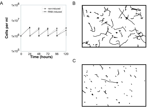
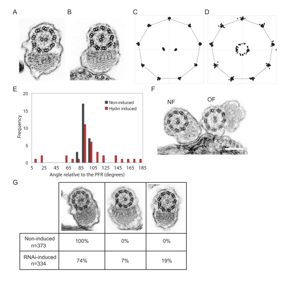
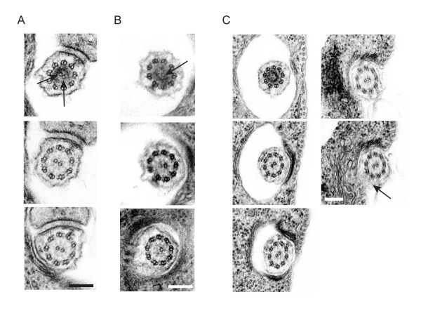
Results: Here we use RNAi in Trypanosoma brucei to address this issue and demonstrate that loss of Hydin causes slow growth and a loss of cell motility. We show that two separate defects in newly-formed flagellar central pair microtubules underlie the loss of cell motility. At early time-points after RNAi induction, the central pair becomes mispositioned, while at later time points the central pair is lost. While the basal body is unaffected, both defects originate at the basal plate, reflecting a role for TbHydin throughout the length of the central pair.

Conclusion: Our data provide the first evidence of Hydin's role within the trypanosome axoneme, and reveal central pair anomalies and thus impairment of ependymal ciliary motility as the likely cause of the hydrocephalus observed in the hy3 mouse.


Figures

Figure 1
Figure 1
TbHydin RNAi-induced cells grow slowly and do not locomote. (A) Representative growth curve shows growth defect starting 48 h after RNAi induction in TbHydin RNAi-induced cells (black squares) compared to non-induced controls (triangles). Cells were maintained in log phase by diluting the culture every 24 h. (B and C) Trajectories of individual cells over the course of a 40 s time-lapse. While non-induced control cells (B) are motile, cells locomotion is severely compromised in TbHydin RNA-induced cells (C).
Figure 2
Figure 2
Central pair microtubule defects in TbHydin RNA-induced cells. (A and B) Transmission electron microscopy images of TbHydin non-induced cells (A) and RNAi-induced cells 48 h after induction (B). Note the aberrant position of the central pair microtubules in (B) compared to the non-induced control. (C-E) Graphical representation of the central pair mispositioning in TbHydin RNAi-induced flagellar profiles (D and red bars in E) compared to the fixed central pair position seen in non-induced controls (C and black bars in E). (F) A cell late on in the cell cycle with two flagella. The new flagellum (NF) has a misoriented central pair while the old flagellum (OF) is normal. (G) Loss of the central pair microtubules in TbHydin RNA-induced cells. Flagellar profiles were analysed at 72 h after RNAi induction, and loss of one or both of the central pair microtubules quantified. Scale bars = 200 nm.
Figure 3
Figure 3
Central pair defects originate at the basal plate. (A-C) Serial thin section electron micrographs through the basal plate region of non-induced (A) and TbHydin RNAi-induced (B and C) cells. In non-induced cells, the two central pair tubules are nucleated simultaneously (A, arrows), however only a single central pair tubule is present in the TbHydin RNA-induced cell (B, arrow). (C) serial thin sectioning of TbHydin RNA-induced cells from the basal plate through to where the flagellum exits the flagellar pocket demonstrates that the central pair mispositioning defect also originates at the basal plate. Central pair position is determined relative to the PFR (arrow). Note how this position is invariant throughout the series. Scale bars = 200 nm.

References

    1. Banizs B, Pike MM, Millican CL, Ferguson WB, Komlosi P, Sheetz J, Bell PD, Schwiebert EM, Yoder BK. Dysfunctional cilia lead to altered ependyma and choroid plexus function, and result in the formation of hydrocephalus. Development. 2005;132:5329–5339. doi: 10.1242/dev.02153. - DOI - PubMed
    1. Chen J, Knowles HJ, Hebert JL, Hackett BP. Mutation of the mouse hepatocyte nuclear factor/forkhead homologue 4 gene results in an absence of cilia and random left-right asymmetry. J Clin Invest. 1998;102:1077–1082. - PMC - PubMed
    1. Ibanez-Tallon I, Gorokhova S, Heintz N. Loss of function of axonemal dynein Mdnah5 causes primary ciliary dyskinesia and hydrocephalus. Hum Mol Genet. 2002;11:715–721. doi: 10.1093/hmg/11.6.715. - DOI - PubMed
    1. Ibanez-Tallon I, Pagenstecher A, Fliegauf M, Olbrich H, Kispert A, Ketelsen UP, North A, Heintz N, Omran H. Dysfunction of axonemal dynein heavy chain Mdnah5 inhibits ependymal flow and reveals a novel mechanism for hydrocephalus formation. Hum Mol Genet. 2004;13:2133–2141. doi: 10.1093/hmg/ddh219. - DOI - PubMed
    1. Sapiro R, Kostetskii I, Olds-Clarke P, Gerton GL, Radice GL, Strauss IJ. Male infertility, impaired sperm motility, and hydrocephalus in mice deficient in sperm-associated antigen 6. Mol Cell Biol. 2002;22:6298–6305. doi: 10.1128/MCB.22.17.6298-6305.2002. - DOI - PMC - PubMed

Publication types