

Long-term cultured human umbilical cord neural-like cells transplanted into the striatum of NOD SCID mice
- PMID: 17683802
- PMCID: PMC2680127
- DOI: 10.1016/j.brainresbull.2007.06.015
Long-term cultured human umbilical cord neural-like cells transplanted into the striatum of NOD SCID mice
Abstract
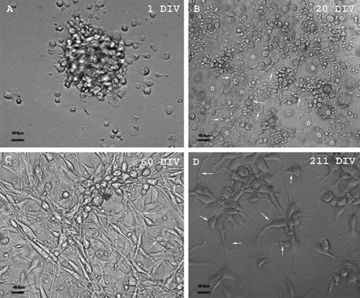
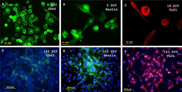
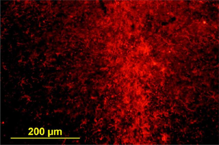
The use of stem cells and other cells as therapies is still in its infancy. One major setback is the limited survival of the grafts, possibly due to immune rejection. Studies were therefore performed with human umbilical cord blood cells (HUCB) to determine the ability of these cells to survive in vivo and the effect of the immune response on their survival by transplantation into the normal striatum of immunodeficient NOD SCID mice. Long-term culture of HUCB cells resulted in several different populations of cells, including one that possessed fine processes and cell bodies that resembled neurons. Their neuronal phenotype was confirmed by immunohistochemical staining for the early neuronal marker TuJ1 and the potentially neural marker Nestin. Five days after cell transplantation of this neuronal phenotype, immunohistochemical staining for human mitochondria confirmed the presence of living HUCB cells in the mouse striatum, with cells localized at the site of injection, expressing early neural and neuronal markers (Nestin and TuJ1) as well as exhibiting neuronal morphology. However, no evidence of surviving cells was apparent 1 month postgrafting. The absence of signs of T cell-mediated rejection, such as CD4 and CD8 lymphocytes and minimal changes in microglia and astrocytes, suggest that cell loss was not due to a T cell-mediated immune response. In conclusion HUCB cells can survive long-term in vitro and undergo neuron-like differentiation. In mice, these cells do not survive a month. This may relate to the differentiated state of the cells transplanted into the unlesioned striatum, rather than T cell-mediated immunological rejection.
Figures


Similar articles
-
Human umbilical cord blood cells express neural antigens after transplantation into the developing rat brain.Cell Transplant. 2002;11(3):265-74. Cell Transplant. 2002. PMID: 12075992
-
Transplantation of human umbilical cord blood-derived adherent progenitors into the developing rodent brain.J Neuropathol Exp Neurol. 2005 Aug;64(8):681-8. doi: 10.1097/01.jnen.0000173892.24800.03. J Neuropathol Exp Neurol. 2005. PMID: 16106216
-
Do hematopoietic cells exposed to a neurogenic environment mimic properties of endogenous neural precursors?J Neurosci Res. 2004 Apr 15;76(2):244-54. doi: 10.1002/jnr.20042. J Neurosci Res. 2004. PMID: 15048922 Free PMC article.
-
[Post-transplant cell immune reconstitution in humanized NOD/SCID mice].Xi Bao Yu Fen Zi Mian Yi Xue Za Zhi. 2014 May;30(5):533-6. Xi Bao Yu Fen Zi Mian Yi Xue Za Zhi. 2014. PMID: 24796750 Chinese.
-
Human cord blood stem cells generate human cytokeratin 18-negative hepatocyte-like cells in injured mouse liver.Am J Pathol. 2005 Aug;167(2):555-64. doi: 10.1016/S0002-9440(10)62997-5. Am J Pathol. 2005. PMID: 16049339 Free PMC article.
Cited by
-
Release activation of iron oxide nanoparticles: (REACTION) a novel environmentally sensitive MRI paradigm.Magn Reson Med. 2011 May;65(5):1253-9. doi: 10.1002/mrm.22839. Epub 2011 Feb 28. Magn Reson Med. 2011. PMID: 21360745 Free PMC article.
-
Potential role of stem cells in severe spinal cord injury: current perspectives and clinical data.Stem Cells Cloning. 2012 Sep 25;5:15-27. doi: 10.2147/SCCAA.S28477. Stem Cells Cloning. 2012. PMID: 24198535 Free PMC article. Review.
-
Neurological disorders and the potential role for stem cells as a therapy.Br Med Bull. 2012;101(1):163-81. doi: 10.1093/bmb/lds001. Epub 2012 Feb 21. Br Med Bull. 2012. PMID: 22357552 Free PMC article. Review.
-
Transplantation of Human Cortically-Specified Neuroepithelial Progenitor Cells Leads to Improved Functional Outcomes in a Mouse Model of Stroke.Front Cell Neurosci. 2021 Apr 29;15:654290. doi: 10.3389/fncel.2021.654290. eCollection 2021. Front Cell Neurosci. 2021. PMID: 33994947 Free PMC article.
-
Human umbilical cord blood-derived mesenchymal stem cell transplantation for the treatment of spinal cord injury.Exp Ther Med. 2014 May;7(5):1233-1236. doi: 10.3892/etm.2014.1608. Epub 2014 Mar 6. Exp Ther Med. 2014. PMID: 24940417 Free PMC article.
References
-
- Bonn D. First cell transplant aimed to reverse stroke damage. Lancet. 1998;352:119. - PubMed
-
- Bouhon IA, Kato H, Chandran S, Allen ND. Neural differentiation of mouse embryonic stem cells in chemically defined medium. Brain Res. Bull. 2005;68:62–75. (Epub September 29, 2005) - PubMed
-
- Buzanska L, Jurga M, Stachowiak EK, Stachowiak MK, Domanska-Janik K. Neural stem-like cell line derived from a nonhematopoietic population of human umbilical cord blood. Stem Cells Dev. 2006;15:391–406. - PubMed
Publication types
MeSH terms
Substances
Grants and funding
LinkOut - more resources
Full Text Sources
Research Materials
