. 2007 Nov;5(11):1282-90; quiz 1245.
doi: 10.1016/j.cgh.2007.05.013. Epub 2007 Aug 1.

Adult autoimmune enteropathy: Mayo Clinic Rochester experience

Affiliations

Adult autoimmune enteropathy: Mayo Clinic Rochester experience

Salma Akram et al. Clin Gastroenterol Hepatol. 2007 Nov.

Abstract

Background & aims: Autoimmune enteropathy is a rare cause of intractable diarrhea associated with circulating gut autoantibodies and a predisposition to autoimmunity. It is rarely observed in adults, with only 11 cases reported to date.

Methods: Fifteen adults with autoimmune enteropathy were identified at the Mayo Clinic, Rochester, from May 2001-June 2006. The demographic, clinical, and treatment data were abstracted from their records.

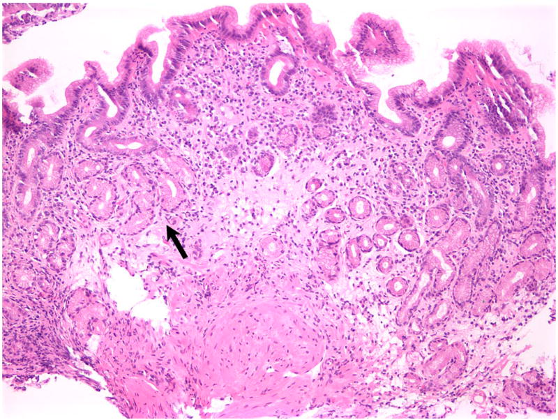
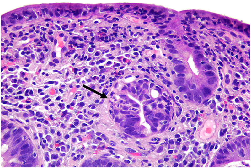
Results: The study population was 87% white, 47% female, with median age of 55 years (interquartile range, 42-67 years). All patients had protracted diarrhea, weight loss, and malnutrition. Celiac disease was excluded by lack of response to gluten-free diet or absence of the celiac disease susceptibility HLA genotypes. Fourteen patients were tested for gut epithelial cell antibodies, and 93% were positive for anti-enterocyte and/or anti-goblet cell antibodies. Predisposition to autoimmune diseases was noted in 80%, as indicated by a variety of circulating autoantibodies. Small intestinal histopathologic findings included subtotal villous atrophy and lymphoplasmacytic infiltration in the lamina propria with relatively few surface intraepithelial lymphocytes. T-cell receptor gene rearrangement studies were negative in all cases. Immunosuppressive therapy was required in 93% of cases. Clinical improvement was noted in 60% after 1-8 weeks of steroid therapy.

Conclusions: Autoimmune enteropathy is a heterogeneous disease and should be considered in the differential diagnosis of malabsorption and small bowel villous atrophy. The presence of gut epithelial cell antibodies can help confirm the diagnosis. No single agent is unequivocally effective in inducing remission, and immunosuppressive therapy is required in most cases.


Figures

Figure 1
Figure 1. Anti-enterocyte antibodies
Indirect immunofluorescence using the patient’s serum on frozen section of normal human small bowel shows a linear fluorescence pattern along the brush border of the enterocytes (× 300).
Figure 2
Figure 2. Anti-goblet cell antibodies
Immunofluorescence staining in the goblet cells mucus, sparing the enterocytes (× 100)
Figure 3
Figure 3
Histopathologic findings in a patient with autoimmune enteritis associated with anti-goblet cell antibodies. (A) Low-power view of the jejunum shows near-total villous atrophy and dense lymphoplasmacellular inflammation in the lamina propria. (B) At higher power, there is surface epithelial injury with disruption of the brush border (arrow) and intense neutrophilic inflammation including crypt abscesses (asterisk). There is no abnormal lymphocytosis in the surface epithelium.
Figure 3
Figure 3
Histopathologic findings in a patient with autoimmune enteritis associated with anti-goblet cell antibodies. (A) Low-power view of the jejunum shows near-total villous atrophy and dense lymphoplasmacellular inflammation in the lamina propria. (B) At higher power, there is surface epithelial injury with disruption of the brush border (arrow) and intense neutrophilic inflammation including crypt abscesses (asterisk). There is no abnormal lymphocytosis in the surface epithelium.
Figure 4
Figure 4
Histopathologic findings in a patient with anti-goblet cell antibodies and pan-gastrointestinal autoimmune disease. (A) Duodenal biopsy demonstrates moderate villous blunting and absence of goblet cells; there is no abnormal surface lymphocytosis. (B) Colonic biopsy with absent goblet cells, mild lymphocytosis in the deep crypt epithelium (arrow), and crypt apoptosis (arrowhead). (C) Gastric biopsy taken from the fundus demonstrates features of autoimmune atrophic gastritis, with complete absence of oxyntic glands and partial replacement by pseudo-pyloric metaplasia (arrow).
Figure 4
Figure 4
Histopathologic findings in a patient with anti-goblet cell antibodies and pan-gastrointestinal autoimmune disease. (A) Duodenal biopsy demonstrates moderate villous blunting and absence of goblet cells; there is no abnormal surface lymphocytosis. (B) Colonic biopsy with absent goblet cells, mild lymphocytosis in the deep crypt epithelium (arrow), and crypt apoptosis (arrowhead). (C) Gastric biopsy taken from the fundus demonstrates features of autoimmune atrophic gastritis, with complete absence of oxyntic glands and partial replacement by pseudo-pyloric metaplasia (arrow).
Figure 5
Figure 5
Histopathologic features in overlap between autoimmune enteropathy and celiac disease. (A) The duodenal biopsy shows near-total villous blunting with crypt architectural distortion including crypt branching (arrow) and crypt destruction (arrowhead), both unusual features of “pure” celiac disease. (B) At high power, there is intense surface intraepithelial lymphocytosis typical for celiac disease, but there is also neutrophilic cryptitis (arrow) and scattered neutrophils in the surface epithelium. This patient had positive anti-enterocyte antibodies, HLA type DQ2, and a weakly positive IgA tissue-transglutaminase antibody.
Figure 5
Figure 5
Histopathologic features in overlap between autoimmune enteropathy and celiac disease. (A) The duodenal biopsy shows near-total villous blunting with crypt architectural distortion including crypt branching (arrow) and crypt destruction (arrowhead), both unusual features of “pure” celiac disease. (B) At high power, there is intense surface intraepithelial lymphocytosis typical for celiac disease, but there is also neutrophilic cryptitis (arrow) and scattered neutrophils in the surface epithelium. This patient had positive anti-enterocyte antibodies, HLA type DQ2, and a weakly positive IgA tissue-transglutaminase antibody.

Similar articles

Cited by

References

    1. Walker-Smith JA, Unsworth DJ, Hutchins P, Phillips AD, Holborow EJ. Autoantibodies against gut epithelium in child with small-intestinal enteropathy. Lancet. 1982;1:566–7. - PubMed
    1. Unsworth DJ, Walker-Smith JA. Autoimmunity in diarrhoeal disease. J Pediatr Gastroenterol Nutr. 41985:375–80. - PubMed
    1. Ruemmele FM, Brousse N, Goulet O. Autoimmune enteropathy: molecular concepts. Curr Opin Gastroenterol. 2004;20:587–91. - PubMed
    1. Biagi F, Corazza GR. Defining gluten refractory enteropathy. Eur J Gastroenterol Hepatol. 2001;13:561–5. - PubMed
    1. Bousvaros A, Leichtner AM, Book L, Shigeoka A, Bilodeau J, Semeao E, Ruchelli E, Mulberg AE. Treatment of pediatric autoimmune enteropathy with tacrolimus (FK506) Gastroenterology. 1996;111:237–43. - PubMed

Publication types

MeSH terms