

EF24 induces G2/M arrest and apoptosis in cisplatin-resistant human ovarian cancer cells by increasing PTEN expression
- PMID: 17684018
- PMCID: PMC4610350
- DOI: 10.1074/jbc.M703796200
EF24 induces G2/M arrest and apoptosis in cisplatin-resistant human ovarian cancer cells by increasing PTEN expression
Abstract
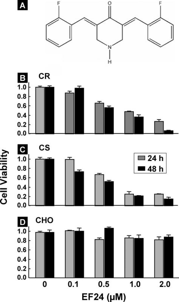
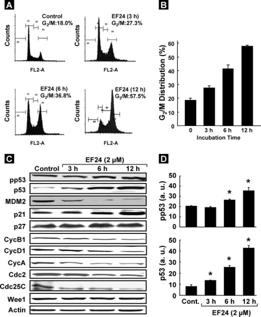
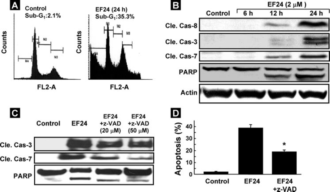
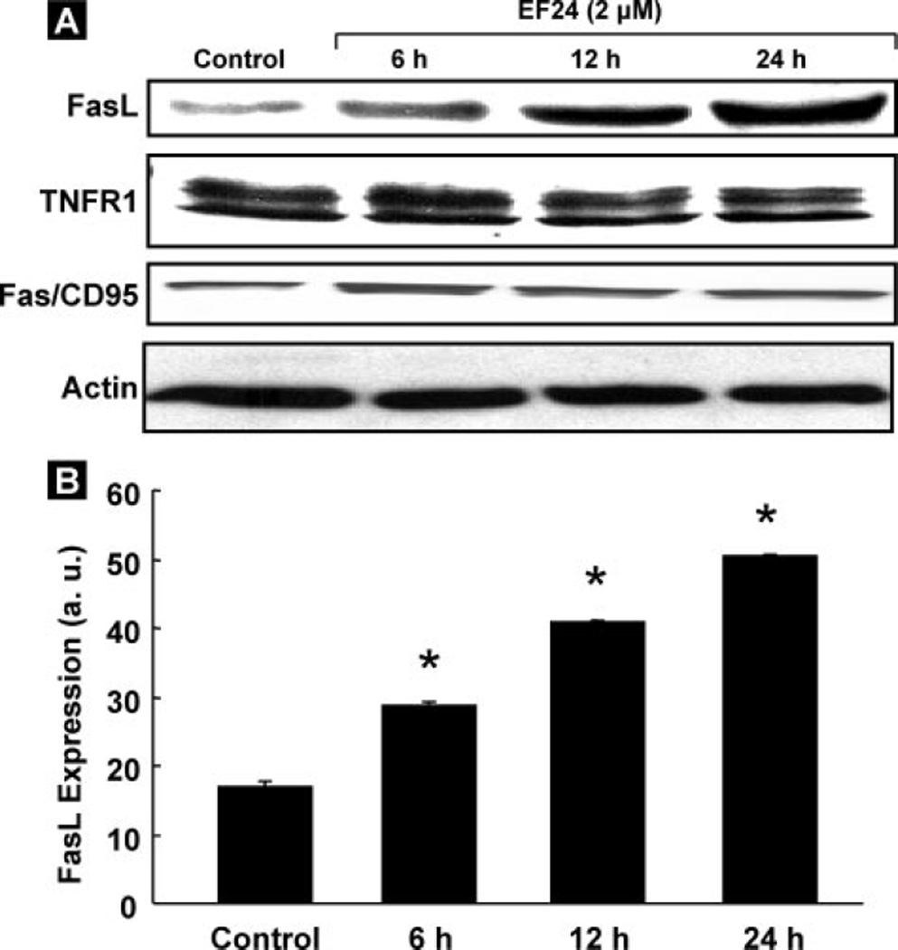
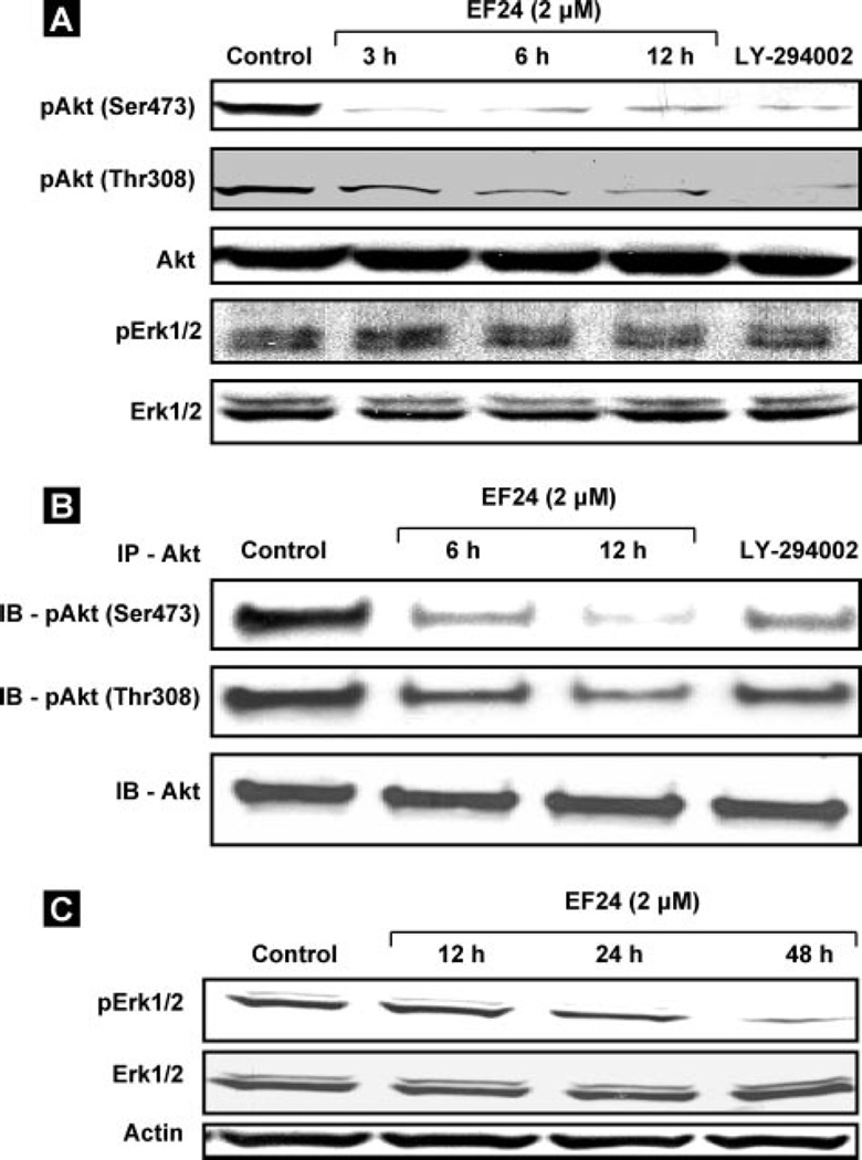
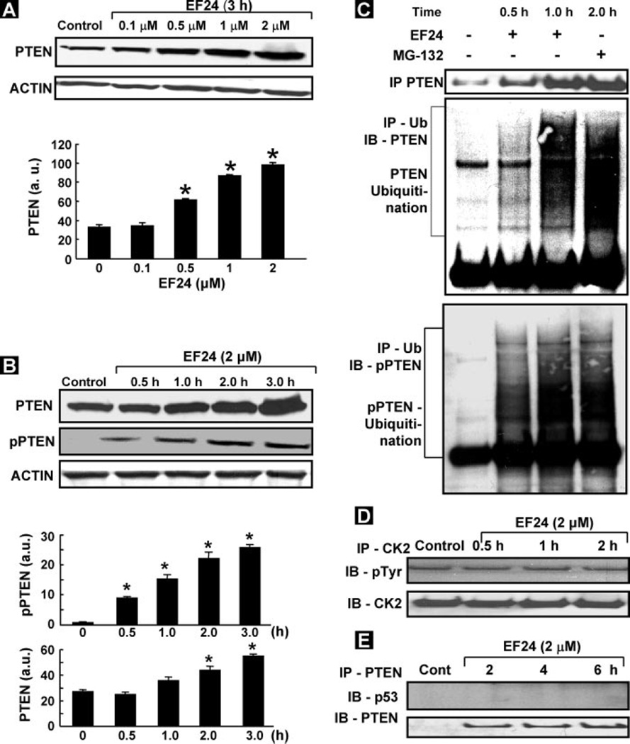
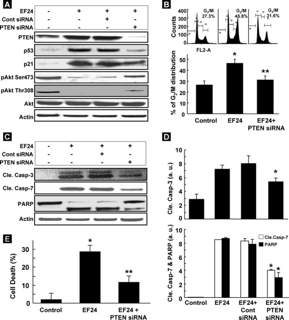
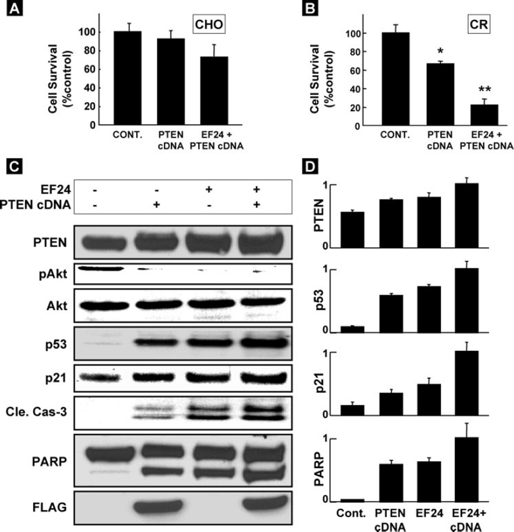
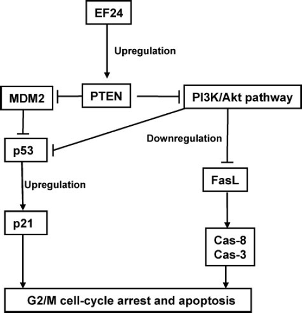
We report that EF24, a synthetic compound 3,5-bis(2-flurobenzylidene)piperidin-4-one, greatly inhibits cisplatin-resistant (CR) human ovarian cancer cell proliferation. The inhibitory effect of EF24 on cell proliferation is associated with G(2)/M phase cell cycle arrest and increased G(2)/M checkpoint protein (pp53, p53, and p21) levels. Within 24 h following treatment, EF24 induced apoptosis in CR cells. The apoptosis was partially blocked by the general caspase inhibitor z-VAD. Within 12 h, EF24 induced a membranous FasL expression, consistent with a substantial decrease in the Ser(473) and Thr(308) phosphorylation of Akt, a known negative regulator of FasL transcription. Also, EF24 activated the phosphorylated PTEN and marginally up-regulated total PTEN expression through the inhibition of ubiquitin-mediated PTEN degradation. Suppression of PTEN expression with siRNA significantly reduced the p53 and p21 levels and activated Akt phosphorylation at Ser(473) and Thr(308), resulting in decreased apoptosis and increased cell survival. On the other hand, overexpression of PTEN markedly induced apoptosis. Our results clearly suggested that EF24 induced significant increase in PTEN expression. The up-regulation of PTEN inhibited Akt and MDM2, which enhanced the level of p53, thereby inducing G(2)/M arrest and apoptosis. Therefore, EF24 appears to have a potential therapeutic role in human ovarian cancer through the activation of PTEN.
Figures
Similar articles
-
In vivo and in vitro suppression of hepatocellular carcinoma by EF24, a curcumin analog.PLoS One. 2012;7(10):e48075. doi: 10.1371/journal.pone.0048075. Epub 2012 Oct 31. PLoS One. 2012. PMID: 23118928 Free PMC article.
-
PTEN- and p53-mediated apoptosis and cell cycle arrest by FTY720 in gastric cancer cells and nude mice.J Cell Biochem. 2010 Sep 1;111(1):218-28. doi: 10.1002/jcb.22691. J Cell Biochem. 2010. PMID: 20506484
-
Over-expression of PTEN sensitizes human ovarian cancer cells to cisplatin-induced apoptosis in a p53-dependent manner.Gynecol Oncol. 2006 Aug;102(2):348-55. doi: 10.1016/j.ygyno.2005.12.033. Epub 2006 Mar 20. Gynecol Oncol. 2006. PMID: 16545436
-
Thymoquinone up-regulates PTEN expression and induces apoptosis in doxorubicin-resistant human breast cancer cells.Mutat Res. 2011 Jan 10;706(1-2):28-35. doi: 10.1016/j.mrfmmm.2010.10.007. Epub 2010 Oct 30. Mutat Res. 2011. PMID: 21040738 Free PMC article.
-
Cisplatin-induced caspase activation mediates PTEN cleavage in ovarian cancer cells: a potential mechanism of chemoresistance.BMC Cancer. 2013 May 10;13:233. doi: 10.1186/1471-2407-13-233. BMC Cancer. 2013. PMID: 23663432 Free PMC article.
Cited by
-
Synergistic antitumor activity of rapamycin and EF24 via increasing ROS for the treatment of gastric cancer.Redox Biol. 2016 Dec;10:78-89. doi: 10.1016/j.redox.2016.09.006. Epub 2016 Sep 21. Redox Biol. 2016. PMID: 27697670 Free PMC article.
-
Therapeutic Inducers of Apoptosis in Ovarian Cancer.Cancers (Basel). 2019 Nov 13;11(11):1786. doi: 10.3390/cancers11111786. Cancers (Basel). 2019. PMID: 31766284 Free PMC article. Review.
-
Autophagy and apoptosis in hepatocellular carcinoma induced by EF25-(GSH)2: a novel curcumin analog.PLoS One. 2014 Sep 30;9(9):e107876. doi: 10.1371/journal.pone.0107876. eCollection 2014. PLoS One. 2014. PMID: 25268357 Free PMC article.
-
Potent Cytotoxicity of Four Cameroonian Plant Extracts on Different Cancer Cell Lines.Pharmaceuticals (Basel). 2020 Oct 31;13(11):357. doi: 10.3390/ph13110357. Pharmaceuticals (Basel). 2020. PMID: 33142782 Free PMC article.
-
Safe and targeted anticancer efficacy of a novel class of antioxidant-conjugated difluorodiarylidenyl piperidones: differential cytotoxicity in healthy and cancer cells.Free Radic Biol Med. 2010 May 1;48(9):1228-35. doi: 10.1016/j.freeradbiomed.2010.02.009. Epub 2010 Feb 12. Free Radic Biol Med. 2010. PMID: 20156552 Free PMC article.
References
-
- Yang X, Fraser M, Moll UM, Basak A, Tsang BK. Cancer Res. 2006;66:3126–3136. - PubMed
-
- Markman M, Howell SB, Lucas WE, Pfeifle CE, Green MR. J. Clin. Oncol. 1984;2:1321–1326. - PubMed
-
- Johnson SW, Ozols RF, Hamilton TC. Cancer. 1993;71(Suppl. 2):644–649. - PubMed
-
- Lee S, Choi EJ, Jin C, Kim DH. Gynecol. Oncol. 2005;97:26–34. - PubMed
Publication types
MeSH terms
Substances
Grants and funding
LinkOut - more resources
Full Text Sources
Other Literature Sources
Medical
Molecular Biology Databases
Research Materials
Miscellaneous
