. 2007 Oct;13(10):1675-92.
doi: 10.1261/rna.594207. Epub 2007 Aug 7.

Nuclear factors are involved in hepatitis C virus RNA replication

Affiliations

Nuclear factors are involved in hepatitis C virus RNA replication

Olaf Isken et al. RNA. 2007 Oct.

Abstract

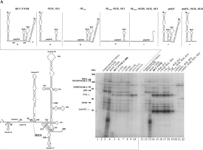
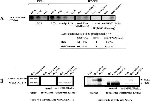
Unraveling the molecular basis of the life cycle of hepatitis C virus (HCV), a prevalent agent of human liver disease, entails the identification of cell-encoded factors that participate in the replication of the viral RNA genome. This study provides evidence that the so-called NF/NFAR proteins, namely, NF90/NFAR-1, NF110/NFAR-2, NF45, and RNA helicase A (RHA), which mostly belong to the dsRBM protein family, are involved in the HCV RNA replication process. NF/NFAR proteins were shown to specifically bind to replication signals in the HCV genomic 5' and 3' termini and to promote the formation of a looplike structure of the viral RNA. In cells containing replicating HCV RNA, the generally nuclear NF/NFAR proteins accumulate in the cytoplasmic viral replication complexes, and the prototype NFAR protein, NF90/NFAR-1, stably interacts with a viral protein. HCV replication was inhibited in cells where RNAi depleted RHA from the cytoplasm. Likewise, HCV replication was hindered in cells that contained another NF/NFAR protein recruiting virus. The recruitment of NF/NFAR proteins by HCV is assumed to serve two major purposes: to support 5'-3' interactions of the viral RNA for the coordination of viral protein and RNA synthesis and to weaken host-defense mechanisms.


Figures

FIGURE 1.
FIGURE 1.
HCV and BVDV RNAs. Organization of HCV and BVDV genomes and replicons. The depicted mono- and bicistronic HCV replicon constructs were applied throughout this work; the bicistronic replicons encoded an additional G418 resistance (NEO) gene (for details, see Grassmann et al. (2005). NTRs are shown as lines, genetic units as shaded boxes, known functions of the viral proteins are indicated (Lindenbach et al. 2007). Proteolytic cleavage sites in the polyprotein are marked as follows: arrow, cleavage by NS3/NS4A; circle, cleavage by signal peptidases; A, autoprotease activity; ubi, cleavage by ubiquitin carboxy hydrolase.
FIGURE 2.
FIGURE 2.
NFAR proteins associate specifically with the HCV 5′- and 3′NTR. (A) Schematic representation of HCV RNA transcripts that were applied in the described affinity purification and UV cross-linking procedures, respectively. (B) Silver-stained SDS-PAGE of affinity-purified proteins: lane 1, control purification without RNA; lane 2, control with 5′NTR-sORF-3′NTR; lane 3, purification performed with TobA-5′NTR-sORF-3′NTR. The bands that contained RHA, NF90/NFAR-1, nucleolin, PTB, NF45, and hnRNP C (all proteins were not present in the controls) are marked. (C) Side-by-side UV cross-linking/competition and Western blot experiments. Cross-linking was performed with BVDV as well as HCV 5′- and 3′NTR transcripts and with non-HCV and HCV non-NTR control transcripts (for details, see Materials and Methods). Left panel: UV cross-linking/competition experiments that were carried out with the HCV 5′NTR transcript and cytoplasmic extracts of Huh-7 and HeLa cells, respectively. For comparison, cross-linking/label transfer experiments with the BVDV 5′- and 3′NTR were separated on the same gel; the supposed protein bands of p130, RHA, NF110/NFAR-2, NF90/NFAR-1, p64, NF45, PTB, and hnRNPC are indicated. Competition was carried out with the indicated molar excess (10× = 10 fold; 100× = 100 fold) of nonlabeled competitor RNA transcript. Right panel: Analogous UV cross-linking/competition experiments carried out with the HCV 3′NTR transcript. Right: for orientation, a Western blot was performed with total protein of HeLa S10 extract and a combination of anti NF45 and anti NF90/NFAR-1 antisera. The Western blot was performed on the very same SDS gel as the cross-linking experiments shown on the left. Extreme right: UV cross-linking/label-transfer experiment performed with HeLa S10 extract and non-NTR and non-HCV (Isken et al. 2003) control transcripts. In the case of the non-NTR control, an RNA transcript corresponding to nucleotides 7891–8162 of Con1 RNA was used (note that experiments with other non-NTR HCV probes led to identical results, not shown).
FIGURE 3.
FIGURE 3.
Mapping of the NFAR protein binding sites in the HCV NTRs. (A) UV cross-linking/label-transfer experiments with mutant transcripts of the HCV 5′- and HCV 3′NTR. (Bottom, left) Scheme of the structure of the HCV (strain 1b) 5′NTR; sites that were used to introduce deletions are indicated (SDS-PAGE); lanes 1–10, cross-linking experiments that were performed with HCV 5′NTR wild-type and mutant (Δ) transcripts (BVDV 5′- and 3′NTR applied as positive controls). The protein bands corresponding to the NFAR proteins, PTB, and hnRNPC are indicated (see also Fig. 2). (Top) Schematic representations of the structures of the wild-type and applied mutant HCV (strain 1b) 3′NTR transcripts (calculated by mfold). (SDS-PAGE) Lanes 11–22, cross-linking experiments performed with the HCV 3′NTR wild-type and mutant transcripts (BVDV 5′- and 3′NTR applied as positive controls). (B) Scheme of the NFAR protein-binding sites in the HCV and BVDV NTRs. The depicted RNA structures (HCV strain 1B; BVDV strain CP7) were experimentally proven or predicted by mfold (quotation marks indicate unverified motifs). The translational start and stop-codons are boxed; arrows indicate the considered 5′-boundary of the IRES and frames of the 3′V and 3′C regions. The known functions of the different RNA domains during translation and replication are indicated (see also text; question marks indicate preliminary data). Shadowed regions designate the defined NFAR protein-binding sites.
FIGURE 3.
FIGURE 3.
Mapping of the NFAR protein binding sites in the HCV NTRs. (A) UV cross-linking/label-transfer experiments with mutant transcripts of the HCV 5′- and HCV 3′NTR. (Bottom, left) Scheme of the structure of the HCV (strain 1b) 5′NTR; sites that were used to introduce deletions are indicated (SDS-PAGE); lanes 1–10, cross-linking experiments that were performed with HCV 5′NTR wild-type and mutant (Δ) transcripts (BVDV 5′- and 3′NTR applied as positive controls). The protein bands corresponding to the NFAR proteins, PTB, and hnRNPC are indicated (see also Fig. 2). (Top) Schematic representations of the structures of the wild-type and applied mutant HCV (strain 1b) 3′NTR transcripts (calculated by mfold). (SDS-PAGE) Lanes 11–22, cross-linking experiments performed with the HCV 3′NTR wild-type and mutant transcripts (BVDV 5′- and 3′NTR applied as positive controls). (B) Scheme of the NFAR protein-binding sites in the HCV and BVDV NTRs. The depicted RNA structures (HCV strain 1B; BVDV strain CP7) were experimentally proven or predicted by mfold (quotation marks indicate unverified motifs). The translational start and stop-codons are boxed; arrows indicate the considered 5′-boundary of the IRES and frames of the 3′V and 3′C regions. The known functions of the different RNA domains during translation and replication are indicated (see also text; question marks indicate preliminary data). Shadowed regions designate the defined NFAR protein-binding sites.
FIGURE 4.
FIGURE 4.
Coimmunoprecipitation of HCV NTR transcripts by anti-NF90/NFAR-1 and anti-NF45 antibodies. The IPs were carried out with a non-immune (control) rabbit antiserum, a rabbit anti NF45 antiserum, and a monoclonal anti NF90/NFAR-1 antibody (see also Materials and Methods). S10 extract of Huh-7 cells was applied as a protein source; the extract was supplemented with equal masses (10 ng) of (32P)-labeled transcripts of the wild-type or mutant HCV 5′ and 3′NTR, respectively (the organization of the applied mutant transcripts is depicted in Fig. 3A). (Top) RNA analysis on a 7 M urea, Tris/borate acrylamide gel; lanes 1–4, transcript input (10% of total input shown; due to their lower molecular weights, the mutant transcripts were applied at a slight molar excess); lanes 5–16, immunoprecipitated RNAs. (Bottom) Protein analysis by SDS-PAGE and Western blot: lanes 5–16, the protein samples that correspond to the above RNA precipitation were probed with a combination of rabbit antisera against NF45 and NF90/NFAR-1. Note that these antisera were different from the originally applied precipitating antibodies (see Materials and Methods). The precipitated NF45 and NF90/NFAR-1 and the costained IgG antibody bands are indicated; lane 17, for orientation, a fraction of nuclear extract was loaded and probed by Western blot and anti-NF90 and anti-NF45 antibody.
FIGURE 5.
FIGURE 5.
NFAR proteins mediate RNA 5′–3′ interactions. (A) 5′–3′ coprecipitation assay. (Left) Silver-stained SDS-PAGE of a chromatographic fraction of the purified [NF90/NFAR-1, NF45, RHA] complex (see also Isken et al. [2003]). (Middle) Schematic sketch of the experimental assay. (Right) Autoradiographs of coprecipitation experiments that were supplemented with the indicated proteins (see Materials and Methods; [NF90/NFAR-1, NF45, RHA] complex abbreviated as [NFAR]). The RNA–RNA coprecipitation experiments were performed in all conceivable combinations, i.e., with the (32P)-labeled NTRs alone (lanes 1–4 and 16–18); with the (32P)-labeled NTRs and biotinylated control RNA (lanes 4–7 and 19–22); with (32P)-labeled 5′NTR and biotinylated 5′NTR (lanes 8–11); (32P)-labeled 5′NTR and biotinylated 3′NTR (lanes 12–15); (32P)-labeled 3′NTR and biotinylated 3′NTR (lanes 23–26); (32P)-labeled 3′NTR and biotinylated 5′NTR (lanes 27–30). The percentile of precipitated labeled RNA is indicated. (B) RNA electron micrographs. (Top) Scheme of the applied RNA molecule (see also Materials and Methods) and analytical agarose gel stained with ethidium bromide indicating the formation of RNA duplex. (Middle) Representative EM images of RNA molecules with wild-type NTRs that were supplemented with BSA or with the purified [NF90/NFAR-1, NF45, RHA] complex. The BSA-supplemented RNAs appeared nearly exclusively (∼98%) as ∼300-nm elongated structures, while ∼14% formed looplike structures in the presence of the NFAR complex (as shown). (Bottom) Statistics of images with wild-type and mutant RNAs; the applied mutant RNAs had a deficient NFAR-binding site in the 3′NTR (−poly-U; see Fig. 3A).
FIGURE 6.
FIGURE 6.
NF90/NFAR-1 interacts with the viral RNA and NS5A in HCV-transfected cells. (A) Coimmunoprecipitation (IP) of NF90/NFAR-1 and HCV RNA. The IP was carried out with anti NF90/NFAR-1 mAb, a control mAb and S10 extracts of 3 × 10⁶ naive or HCV replicon-transfected Huh-7 cells. Coprecipitated viral RNA was detected by RT-PCR, which generated a 270-bp product. As controls and for semiquantification, we performed PCRs or RT-PCRs with the indicated amounts of cDNA, replicon transcript, and total RNA extracted from 3 × 10⁴ naive or HCV replicon-transfected cells. The amount of HCV RNA that was coprecipitated by the anti NFAR-1 antibody and by the control mAb was estimated with respect to these controls (considering that these controls applied a 100-fold lower amount of cells): the table shows average values of four experiments. Note that precipitations that were carried out in the presence of micrococcal nuclease (75 U/extract of 106 cells) resulted negative results (not shown). (B) Co-IP of NF90/NFAR-1 and NS5A. The IP was performed with anti NF90/NFAR-1 mAb and a control mAb using RNase-treated S10 extracts of naive Huh-7 (Huh), Huh-7 that carried HCV replicon (Huh/replicon), and Huh-7 that had been transfected with a plasmid expressing the HCV NS5A (Huh/NS5A). Precipitated NF90/NFAR-1 was detected by Western blots with an antiserum that also detects NF110/NFAR-2 (see control with total, untreated extract; note that the anti-NF90/NFAR-1 mAb applied for IP did not coprecipitate NF110). Expression and coprecipitation of NS5A was detected by Western blots with a rabbit serum against the viral protein (staining NS5A in each case as a double band). Note that the control antibody yielded a stronger IgG band than the NF90/NFAR-1 mAb. Co-IP of NF90/NFAR-1 and NS5A was detected irrespective of whether the extract was treated with RNase A (20 μg/extract of 106 cells) or not.
FIGURE 7.
FIGURE 7.
NF/NFAR proteins accumulate at the sites of viral replication in the cytoplasm. Representative confocal images (600× enlarged) of MDBK cells infected with BVDV and of persistently HCV replicon-transfected Huh-7 cells. (Upper panel) MDBK cells were infected at 0.1 MOI with BVDV strain CP7, 24 h post-infection, the cells were stained with an anti BVDV NS3 mAb and counterstained with a rabbit antiserum against NF45. (Lower panels) Huh-7 cells carrying the HCV (Con1, strain 1B) replicon were stained with a mAb against NS5A and counterstained with antisera against the different NFAR proteins. Both fields were independently analyzed, and a colocalization of viral and cellular proteins is shown by merging. (Bottom) HCV replicon-carrying cells stained with anti-NS5A and anti-NF90/NFAR-1 antibody (enlarged).
FIGURE 8.
FIGURE 8.
HCV replication is inhibited in cells with lower levels of RHA. The experiment was carried out with RNAi depleted Huh-7 cells that were transiently transfected with monocistronic HCV replicon RNA (see Materials and Methods). (Upper panel) Western blot indicating the level of RHA at day 4 post-transfection of anti-RHA or control siRNAs (error bars indicate mean deviations of three different experiments. (Lower panel) Representative RNase protection experiment measuring HCV RNA replication at days 2–5 post-transfection in cells that had been transfected with anti RHA or control siRNAs. The same cell preparations were also applied in previous experiments with BVDV; GAPDH was used as a loading control. Note that the cells showed no signs of apoptosis over this time and that replication was equally inhibited when we transfected varying amounts of HCV RNA (Isken et al. [2003] and data not shown).
FIGURE 9.
FIGURE 9.
Interference of HCV and BVDV replication. Naive and HCV replicon carrying Huh-7 cells were transfected with replicative or non-replicative (RdRp inactivated) monocistronic BVDV replicon. Replication of HCV and BVDV was monitored by complementary IF staining of the cells with anti-HCV NS5A rabbit antiserum and anti-BVDV NS3 mAb, respectively. The bar diagram reflects a representative experiment that determined the fractions of cells of a tissue culture plate (the total number of cells was calculated by DAPI staining of the nuclei) that support HCV or BVDV replication in the absence (set 100%) or presence of the respective other RNA. (Right panel) Transfection experiments of naïve and HCV replicon carrying Huh-7 cells with fluorescent-marked (Cy3) siRNAs indicating identical receptiveness of both cell types for RNA transfections.

References

    1. Alvarez, D.E., Lodeiro, M.F., Luduena, S.J., Pietrasanta, L.I., Gamarnik, A.V. Long-range RNA–RNA interactions circularize the dengue virus genome. J. Virol. 2005;79:6631–6643. - PMC - PubMed
    1. Appel, N., Herian, U., Bartenschlager, R. Efficient rescue of hepatitis C virus RNA replication by trans-complementation with nonstructural protein 5A. J. Virol. 2005;79:896–909. - PMC - PubMed
    1. Barton, D.J., Morasco, B.J., Flanegan, J.B. Translating ribosomes inhibit poliovirus negative-strand RNA synthesis. J. Virol. 1999;73:10104–10112. - PMC - PubMed
    1. Becher, P., Orlich, M., Thile, H.J. Mutations in the 5′ nontranslated region of bovine viral diarrhea virus result in altered growth characteristics. J. Virol. 2000;74:7884–7894. - PMC - PubMed
    1. Behrens, S.E., Isken, O. Cis and trans-acting factors in Flaviviridae replication. In: Kalitzky M., Borowski P., editors. Molecular Biology of the Flavivirus. Horizon Bioscience; Norwich, UK: 2006. pp. 101–134.

Publication types