

Role of CD4+ T cells in sarcoidosis
- PMID: 17684290
- PMCID: PMC2647597
- DOI: 10.1513/pats.200606-130MS
Role of CD4+ T cells in sarcoidosis
Abstract
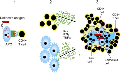
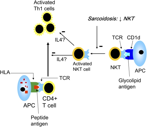
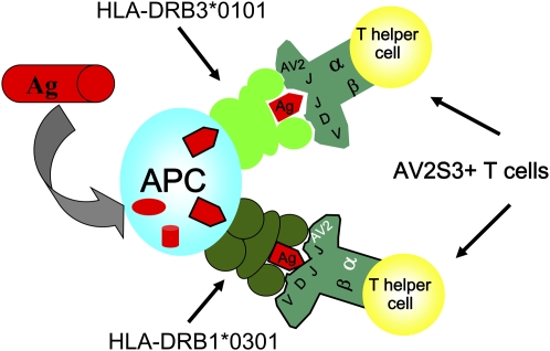
Activated pulmonary CD4(+) T lymphocytes of the Th-1 type are essential for the inflammatory process in sarcoidosis, and IFN-gamma production is crucial for the characteristic granuloma formation. Both the T cells and their inflammatory mediators may constitute possible targets for immunotherapy. A particular T-cell subset, the T-cell receptor (TCR) AV2S3(+) bronchoalveolar lavage (BAL) CD4(+) T cells, is found at dramatically increased levels in the BAL fluid of human leukocyte antigen (HLA)-DRB1*0301-positive and/or HLA-DRB3*0101-positive patients with sarcoidosis. The AV2S3(+) BAL CD4(+) T cells strongly associate with the sarcoid inflammation, and future studies on this particular T-cell subset to reveal their specificity may lead to the identification of sarcoidosis-specific antigen(s). T-cell subpopulations with regulatory functions (i.e., natural killer T cells and T regulatory cells) have recently been described as abnormal in sarcoidosis. Dysfunctional regulatory T cells may allow T effector cells to contribute to the formation of granulomas, and they may thus be relevant for the inflammatory process in this disease. These findings are exciting news and will be of help in designing new treatment strategies.
Figures
References
-
- Hunninghake GW, Crystal RG. Pulmonary sarcoidosis: a disorder mediated by excess helper T-lymphocyte activity at sites of disease activity. N Engl J Med 1981;305:429–434. - PubMed
-
- Pinkston P, Bitterman PB, Crystal RG. Spontaneous release of interleukin-2 by lung T lymphocytes in active pulmonary sarcoidosis. N Engl J Med 1983;308:793–800. - PubMed
-
- ATS/ERS/WASOG. Statement on sarcoidosis: joint statement of the American Thoracic Society (ATS), the European Respiratory Society (ERS), and the World Association of Sarcoidosis and Other Granulomatous Disorders (WASOG) adopted by the ATS Board of Directors and by the ERS Executive Committee, February 1999. Am J Respir Crit Care Med 1999;160:736–755. - PubMed
Publication types
MeSH terms
Grants and funding
LinkOut - more resources
Full Text Sources
Other Literature Sources
Research Materials