. 2007 Aug 8;2(8):e705.
doi: 10.1371/journal.pone.0000705.

The murine gammaherpesvirus-68 gp150 acts as an immunogenic decoy to limit virion neutralization

Affiliations

The murine gammaherpesvirus-68 gp150 acts as an immunogenic decoy to limit virion neutralization

Laurent Gillet et al. PLoS One. .

Abstract

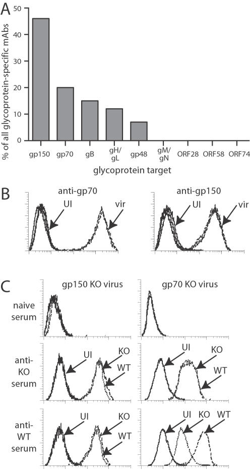
Herpesviruses maintain long-term infectivity without marked antigenic variation. They must therefore evade neutralization by other means. Immune sera block murine gammaherpesvirus-68 (MHV-68) infection of fibroblasts, but fail to block and even enhance its infection of IgG Fc receptor-bearing cells, suggesting that the antibody response to infection is actually poor at ablating virion infectivity completely. Here we analyzed this effect further by quantitating the glycoprotein-specific antibody response of MHV-68 carrier mice. Gp150 was much the commonest glycoprotein target and played a predominant role in driving Fc receptor-dependent infection: when gp150-specific antibodies were boosted, Fc receptor-dependent infection increased; and when gp150-specific antibodies were removed, Fc receptor-dependent infection was largely lost. Neither gp150-specific monoclonal antibodies nor gp150-specific polyclonal sera gave significant virion neutralization. Gp150 therefore acts as an immunogenic decoy, distorting the MHV-68-specific antibody response to promote Fc receptor-dependent infection and so compromise virion neutralization. This immune evasion mechanism may be common to many non-essential herpesvirus glycoproteins.


Conflict of interest statement

Competing Interests: The authors have declared that no competing interests exist.

Figures

Figure 1
Figure 1. Gp150 is the most immunogenic MHV-68 glycoprotein.
A. Hybridomas were derived from the pooled spleens of 3 BALB/c mice 5 months after intranasal MHV-68 infection. Each mAb that recognized unfixed, MHV-68-infected BHK-21 cells was typed for its glycoprotein target as outlined in Table 1. None remained unaccounted for. The data shown are from 1 fusion experiment that generated 128 glycoprotein-specific mAbs. 5 further experiments all gave similar results. B. BHK-21 cells were either uninfected (UI, solid lines) or infected with wild-type MHV-68 (2 PFU/cell, 18h: vir, dashed lines) and stained with mAbs specific for gp70 or gp150. Histograms for 3 mAbs, all used in saturating amounts, are overlaid in each graph. Gp70-specific staining was consistently stronger than that of other viral glycoproteins, including gp150. C. BHK-21 cells were either uninfected (UI, solid lines) or infected (2 PFU/cell, 18h) with wild-type (WT, dashed lines), gp150-deficient (gp150 KO, dotted lines) or gp70-deficient (gp70 KO, dotted lines) viruses. The cells were then stained with sera from mice infected with wild-type, gp150-deficient or gp70-deficient viruses, or from age-matched naive mice.
Figure 2
Figure 2. Epitope mapping for gp150-specific mAbs.
A. Gp150 comprises an N-terminal signal sequence, a small N-terminal domain, a long stalk rich in anionic residues, proline residues and potential O-glycosylation sites (A/D/E/P/T/S-rich domain), a transmembrane domain and a short cytoplasmic tail. Our analysis focussed on the membrane-distal half of the protein. B. GST fusion proteins were purified with glutathione-sepharose, separated by SDS-PAGE and Coomassie stained. Longer regions of the stalk domain were less well expressed than shorter forms and were subject to more degradation. C. MAb recognition was mapped by ELISA with GST fusion proteins covering the gp150 extracellular domain. Examples of fusion protein recognition are shown (mAbs T1A1, T4G2 and 150-41, with their deduced recognition sites given below). Despite the limited amino acid diversity of gp150, little mAb cross-reactivity was evident between the expressed protein segments. The gH/gL-specific mAb T4C5 provided a negative control. D. Recognition patterns were confirmed by flow cytometric staining of 293T cells transfected with either full-length gp150 or the N-terminal 151 residues of gp150 with a GPI membrane anchor. Examples of staining are shown. Control = secondary antibody only. E. More detailed analysis of the N21-C151 region, which accounted for most gp150-specific mAbs, using N-terminal gp150 truncations. Virus = wells coated with 0.01% Triton X-100-disrupted MHV-68 virions, GST = GST alone. Examples of recognition patterns are shown. The gp70-specific mAb 6H10 provided a negative control for gp150 recognition. F. Summary of GST fusion protein recognition by 81 gp150-specific mAbs. G. MAb specificities within the 108-151 region were mapped with overlapping 15-mer biotinylated peptides bound to avidin-coated plates. The amino acid residues covered by each peptide are indicated. Examples of recognition patterns are shown with the total number of mAbs showing that pattern.
Figure 3
Figure 3. Effect of gp150-specific mAbs on MHV-68 virion infectivity.
A. EGFP-expressing MHV-68 virions were incubated with hybridoma supernatants (1 h, 37°C) and then added to RAW264.7 macrophages (3 PFU/cell). 18h later, the number of infected cells was determined by flow cytometric assay of viral eGFP expression. The mAbs are grouped by the region of gp150 recognized. The bars indicate the %eGFP+ RAW264.7 cells in each culture. B. EGFP-expressing MHV-68 virions were incubated (1 h, 37°C) with dilutions of the gp150-specific mAb T4G2 and then added to either BHK-21 fibroblasts or RAW264.7 macrophages. 18h later, the number of infected cells was determined by flow cytometric assay of viral eGFP expression. The data shown are representative of 3 equivalent experiments C. EGFP-expressing MHV-68 virions were incubated with gp150-specific mAbs (T7F5, T1A1), with immune serum, or with no antibody and then added to normal, GAG+ CHO cells or to the GAG-deficient CHO cell mutant pgs-745 (GAG). Infection was quantitated 18h later by flow cytometric assay of viral eGFP expression.
Figure 4
Figure 4. Serum-mediated, FcR-dependent MHV-68 infection depends on gp150.
A. Wild-type or gp150-deficient virions were incubated with sera from BALB/c mice, taken 2 months after infection with wild-type (WT), gp150-deficient (M7-FRT, M7-STOP) or revertant (REV) viruses as indicated. The virus/serum mixtures were then added to RAW264.7 cells (3 PFU/cell). The proportion of infected cells in each culture was determined 18h later by flow cytometric assay of viral eGFP expression. nil = uninfected, vir = virus only. Each serum sample was pooled from 5 mice. The data are from 1 of 2 equivalent experiments. B. Sera were taken from C57BL/6 mice 6 months after infection with either wild-type (WT) or M7-STOP (gp150) viruses. EGFP-expressing WT or gp150 viruses were then incubated with sera from individual mice and used to infect RAW264.7 cells. The level of infection was assayed 18h later by flow cytometry of viral eGFP expression. C. Sera were pooled from 5 mice at 6 months after infection with wild-type (WT) or gp150-deficient (M7-) MHV-68. Gp150-specific antibodies were removed from wild-type-immune sera by 3 rounds of incubation on 2% paraformaldehyde-fixed CHO-gp150 cells (see Figure 2D) (WT abs-150). Controls were incubated on paraformaldehyde-fixed CHO cells (WT abs-C). Wild-type or gp150-deficient viruses were incubated with each serum sample and then used to infected RAW264.7 cells (3 PFU/cell, 18 h). The data are representative of 2 experiments.
Figure 5
Figure 5. The antibody response to gp150 tends to reduce rather than enhance MHV-68 neutralization.
A. Wild-type MHV-68 virions (1000 PFU/sample) were incubated with sera pooled from 5 mice 2 months after infection with gp150+ (WT, REV) or gp150 (M7-FRT, M7-STOP) viruses and then used to infect BHK-21 cells. Control samples were BHK-21 cells infected with virus and no antibody. B. Sera from C57BL/6 mice 3 months after infection with wild-type (WT) or M7-STOP (gp150) MHV-68 was used to inhibit wild-type MHV-68 infection of BHK-21 cells. Despite variation between individual mice, plaque titers were significantly lower with the 2% and 6% sera from gp150 knockout-infected mice (p<0.01 by Student's t test).
Figure 6
Figure 6. gp150-specific immunity in carrier mice increases FcR-dependent infection and has no effect on neutralization.
A. BHK-21 cells were left uninfected (solid lines) or infected (2PFU/cell 18h) with MHV-68 (dashed lines) or VAC-150 (dotted lines), then assayed for cell surface gp150 expression by flow cytometry. MAb T4G2 recognizes an epitope in gp150 amino acid residues 108–151. MAb T1A1 recognizes an epitope in amino acid residues 152–269. VAC-150 expresses residues 1–151 with a GPI anchor. B. BALB/c (BC) or C57BL/6J (B6) mice were either left uninfected or infected with VAC-gp150. At the times post-infection indicated, sera were incubated with eGFP-expressing MHV-68. The serum/virus mixtures were then used to infect RAW264.7 macrophages. Infection was quantitated 18h later by flow cytometric assay of viral eGFP expression. The data are from 1 of 2 equivalent experiments. C. C57BL/6 mice were infected intranasally with MHV-68. 3 months later they were boosted with VAC-150 (+VAC-150) or a control vaccinia virus expressing the Herpes simplex virus gE (+VAC-gE) or left unboosted (MHV-68). Naive = age-matched, uninfected mice. Immune sera were pooled from groups of 3 mice. A and B are separate pools for each treatment arm. The sera were assayed by ELISA for reactivity against N-terminal (residues 21–151) or C-terminal (residues 269–461) fragments of gp150. Only the N-terminal part is expressed by VAC-150. D. The same sera as in C were added to eGFP+ MHV-68 virions and the mixtures added to RAW264.7 cells. 18h later, eGFP expression was quantitated by flow cytometry. The data are from 1 of 3 equivalent experiments. E. C57BL/6 and BALB/c mice were either left uninfected (naive), infected with MHV-68 or VAC-gp150, or infected with MHV-68 and then boosted with VAC-gp150. Pooled sera from these mice (5 per group) were then tested for MHV-68 neutralization by plaque assay. Infection with VAC-gp150 made no detectable difference to neutralization titers. The data are from 1 of 2 equivalent experiments.

Similar articles

Cited by

References

    1. Zinkernagel RM, Hengartner H. Protective ‘immunity’ by pre-existent neutralizing antibody titers and preactivated T cells but not by so-called ‘immunological memory’. Immunol Rev. 2006;211:310–319. - PubMed
    1. Sissons JG, Oldstone MB. Killing of virus-infected cells: the role of antiviral antibody and complement in limiting virus infection. J Infect Dis. 1980;142:442–448. - PubMed
    1. Sitki-Green D, Covington M, Raab-Traub N. Compartmentalization and transmission of multiple Epstein-Barr virus strains in asymptomatic carriers. J Virol. 2003;77:1840–1847. - PMC - PubMed
    1. Xu J, Lyons PA, Carter MD, Booth TW, Davis-Poynter NJ, et al. Assessment of antigenicity and genetic variation of glycoprotein B of murine cytomegalovirus. J Gen Virol. 1996;77:49–59. - PubMed
    1. Roth MG, Compans RW. Antibody-resistant spread of vesicular stomatitis virus infection in cell lines of epithelial origin. J Virol. 1980;35:547–550. - PMC - PubMed

Publication types