

doi: 10.1186/1749-8104-2-15.
Making sense of zebrafish neural development in the Minervois
Affiliations
- PMID: 17686145
- PMCID: PMC1988804
- DOI: 10.1186/1749-8104-2-15
Item in Clipboard
Making sense of zebrafish neural development in the Minervois
Neural Dev.
.
Abstract
The meeting 'From sensory perception to motor output: genetic bases of behavior in the zebrafish embryo' was held at Minerve (South of France) on March 16-18, 2007. The meeting site was beautifully situated in the heart of the Minervois wine country, and its remoteness promoted conversations and interaction over the course of the program. The meeting covered neurogenesis and eye development on day 1, ear and lateral line development on day 2, and brain connectivity and behavior on day 3. Underlying all sessions, however, ran the growing importance of live imaging, an approach that takes full advantage of the transparency of fish embryos and early larvae, as illustrated by several movies and links in this report.
Figures
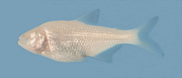
Astyanax fasciatus, cave form. Picture provided by B Jeffery.
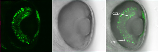
Example of a shh enhancer specific for the ganglion cell layer (GCL) and inner nuclear layer (INL) of the retina, driving expression of GFP (72 hpf embryo carrying the -2.shh:gfpAC11 transgene). Figure provided by Saradavey Rathnam in U Strähle's lab.
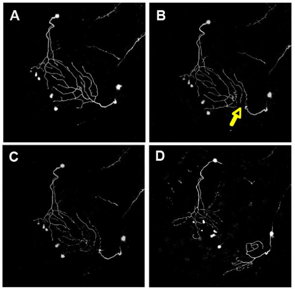
Imaging peripheral arbor re-innervation in vivo. Confocal projections showing dorsal views of two trigeminal axons, visualized with GFP, in a 54 hpf zebrafish embryo at several time points during a two-photon axotomy experiment. Anterior is to the bottom left. Images are each 420 microns across. (a) Twenty minutes before axotomy. (b) Approximately 20 minutes after axotomy. Yellow arrow points to site of axotomy. (c) Two hours after axotomy, the distal portion degenerates. (d) Robust regrowth is apparent 12 hours after axotomy. Figure provided by A Sagasti.
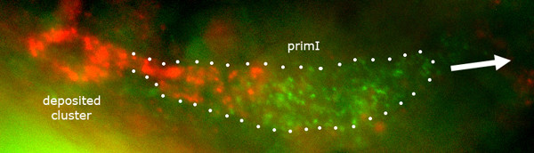
Expression of cxcr4 and cxcr7 in the migrating primordium. Double in situ hybridization with a cxcr4b probe (green) and a cxcr7 probe (red). cxcr7 is exclusively expressed by cells in the trailing region and in cells that are being deposited, while cxcr4b is more ubiquitously expressed (albeit at a higher level by the cells in the leading region). White dots outline the migrating primordium, primI. The direction of migration is shown by the white arrow. Picture provided by C Dambly-Chaudière and M Rossel.
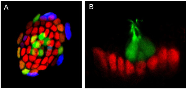
Distribution of Sox2 in lateral line neuromasts. (a) Confocal image of a zebrafish lateral line neuromast. Mantle cells and hair cells are labeled by GFP (green) in this transgenic line of zebrafish. Fluorescent immunostaining reveals expression of the neural progenitor marker protein Sox2 (red) and BrdU incorporation (blue). Cell division is occurring mostly in the periphery of the neuromast. (b) Confocal image of a zebrafish lateral line neuromast inmunostained to detect the neural progenitor marker protein Sox2 (red) and GFP (green) in hair cells. Note that these two markers do not overlap, suggesting that Sox2 is not present in differentiated cell types in neuromasts. Pictures provided by M Allende.
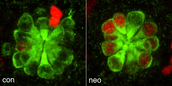
Control (con) and neomycin (neo) treated zebrafish lateral line neuromasts 72 hours after antibiotic treatment. Regenerated hair cells stained with anti-myosin VI antibody (green) are derived from proliferative precursors that incorporated BrdU (red). Figure provided by D Raible.
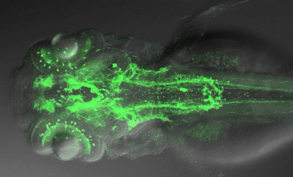
Projection on the horizontal plane of a Z-stack movie shown by J Schweitzer, illustrating a whole mount 3 dpf embryo labeled with an antibody against tyrosine hydroxylase. This picture was generated by S Ryu and W Driever.
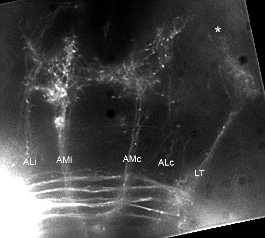
Second-order projection from the lateral line, at the level of rhombomeres 1–2. Two large branches, AMc and AMi, extend symmetrically on either side of the midline towards the oculomotor nuclei and nuclei of the Median Longitudinal Fascicle (MLF). More laterally, two ill-defined branches extend to forebrain nuclei (ALc and ALi). A fifth branch extends to the contralateral torus semicircularis (LT). At the anterior edge of the LT branch, a few fibers extend dorsally (asterisk) into the tectum. Dorsal view, composite of several focal planes. The complete Z-stack is shown as Additional file 7. Figure provided by A Ghysen.
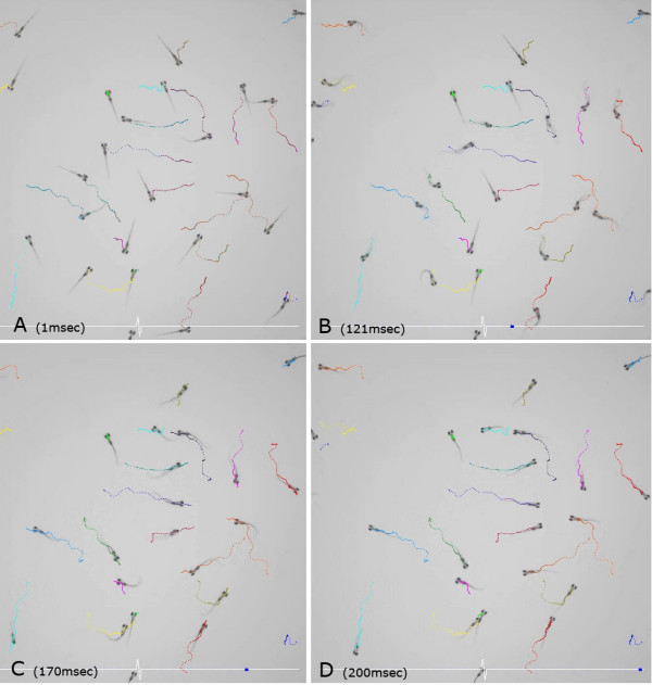
Four still pictures at different stages of the startle response, taken from Additional file 11. The pictures were taken at (a) 1, (b) 121, (c) 170 and (a) 200 ms after stimulation. Figure provided by M Granato.
References
-
- Program of the Minerve Meeting http://raibleweb.biostr.washington.edu/program.html
-
- Pictures of the Meeting http://raibleweb.biostr.washington.edu/minervephotos.html
-
- Timelapse of Eye Morphogenesis http://chien.neuro.utah.edu/media/main.php?g2_itemId=38
-
- Timelapse of Cell Behaviors During Eye Morphogenesis http://chien.neuro.utah.edu/media/main.php?g2_itemId=41
-
- Sagasti Lab Movies http://www.mcdb.ucla.edu/Research/Sagasti/data/axotomyandablation.html
Publication types
MeSH terms
LinkOut - more resources
Full Text Sources
Research Materials
Miscellaneous
