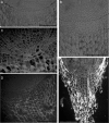
Influence of a weak DC electric field on root meristem architecture
- PMID: 17686761
- PMCID: PMC2749630
- DOI: 10.1093/aob/mcm164
Influence of a weak DC electric field on root meristem architecture
Abstract
Background and aims: Electric fields are an important environmental factor that can influence the development of plants organs. Such a field can either inhibit or stimulate root growth, and may also affect the direction of growth. Many developmental processes directly or indirectly depend upon the activity of the root apical meristem (RAM). The aim of this work was to examine the effects of a weak electric field on the organization of the RAM.
Methods: Roots of Zea mays seedlings, grown in liquid medium, were exposed to DC electric fields of different strengths from 0.5 to 1.5 V cm(-1), with a frequency of 50 Hz, for 3 h. The roots were sampled for anatomical observation immediately after the treatment, and after 24 and 48 h of further undisturbed growth.
Key results: DC fields of 1 and 1.5 V cm(-1) resulted in noticeable changes in the cellular pattern of the RAM. The electric field activated the quiescent centre (QC): the cells of the QC penetrated the root cap junction, disturbing the organization of the closed meristem and changing it temporarily into the open type.
Conclusions: Even a weak electric field disturbs the pattern of cell divisions in plant root meristem. This in turn changes the global organization of the RAM. A field of slightly higher strength also damages root cap initials, terminating their division.
Figures


Similar articles
-
Effect of mechanical stress on Zea root apex. I. Mechanical stress leads to the switch from closed to open meristem organization.J Exp Bot. 2011 Aug;62(13):4583-93. doi: 10.1093/jxb/err169. Epub 2011 Jun 9. J Exp Bot. 2011. PMID: 21659665 Free PMC article.
-
Length and activity of the root apical meristem revealed in vivo by infrared imaging.J Exp Bot. 2015 Mar;66(5):1387-95. doi: 10.1093/jxb/eru488. Epub 2014 Dec 24. J Exp Bot. 2015. PMID: 25540436 Free PMC article.
-
Lateral root development in the maize (Zea mays) lateral rootless1 mutant.Ann Bot. 2013 Jul;112(2):417-28. doi: 10.1093/aob/mct043. Epub 2013 Feb 28. Ann Bot. 2013. PMID: 23456690 Free PMC article.
-
Root apical meristem diversity and the origin of roots: insights from extant lycophytes.J Plant Res. 2020 May;133(3):291-296. doi: 10.1007/s10265-020-01167-2. Epub 2020 Jan 30. J Plant Res. 2020. PMID: 32002717 Review.
-
The organization of roots of dicotyledonous plants and the positions of control points.Ann Bot. 2011 May;107(7):1213-22. doi: 10.1093/aob/mcq229. Epub 2010 Nov 29. Ann Bot. 2011. PMID: 21118839 Free PMC article. Review.
Cited by
-
eSoil: A low-power bioelectronic growth scaffold that enhances crop seedling growth.Proc Natl Acad Sci U S A. 2024 Jan 9;121(2):e2304135120. doi: 10.1073/pnas.2304135120. Epub 2023 Dec 26. Proc Natl Acad Sci U S A. 2024. PMID: 38147542 Free PMC article.
-
Root apex transition zone as oscillatory zone.Front Plant Sci. 2013 Oct 2;4:354. doi: 10.3389/fpls.2013.00354. eCollection 2013. Front Plant Sci. 2013. PMID: 24106493 Free PMC article.
-
Root electrotropism in Arabidopsis does not depend on auxin distribution but requires cytokinin biosynthesis.Plant Physiol. 2022 Mar 4;188(3):1604-1616. doi: 10.1093/plphys/kiab587. Plant Physiol. 2022. PMID: 34893912 Free PMC article.
-
Effect of mechanical stress on Zea root apex. I. Mechanical stress leads to the switch from closed to open meristem organization.J Exp Bot. 2011 Aug;62(13):4583-93. doi: 10.1093/jxb/err169. Epub 2011 Jun 9. J Exp Bot. 2011. PMID: 21659665 Free PMC article.
-
Root Tropisms: Investigations on Earth and in Space to Unravel Plant Growth Direction.Front Plant Sci. 2020 Feb 21;10:1807. doi: 10.3389/fpls.2019.01807. eCollection 2019. Front Plant Sci. 2020. PMID: 32153599 Free PMC article. Review.
References
-
- Barlow PW. Regeneration of the cap of primary roots of Zea mays. New Phytology. 1974;73:937–954.
-
- Barlow PW. The root cap: cell dynamics, cell differentiation and cap function. Journal of Plant Growth Regulation. 2003;21:261–286.
-
- Barlow PW, Luck HB, Luck J. Autoreproductive cells and plant meristem construction: the case of the tomato cap meristem. Protoplasma. 2001;215:50–63. - PubMed
-
- Baum SF, Rost TL. Root apical organization in Arabidopsis thaliana 1: root cap and protoderm. Protoplasma. 1996;192:178–188.
-
- van den Berg C, Willemsen V, Hendriks G, Weisbeek P, Scheres B. Short-range control of cell differentiation in the Arabidopsis root meristem. Nature. 1997;390:287–289. - PubMed
MeSH terms
LinkOut - more resources
Full Text Sources
Miscellaneous

