Stimulation of NEIL2-mediated oxidized base excision repair via YB-1 interaction during oxidative stress
- PMID: 17686777
- PMCID: PMC2679419
- DOI: 10.1074/jbc.M704672200
Stimulation of NEIL2-mediated oxidized base excision repair via YB-1 interaction during oxidative stress
Abstract
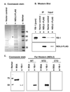
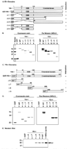
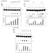
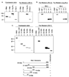
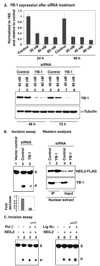
The recently characterized enzyme NEIL2 (Nei-like-2), one of the four oxidized base-specific DNA glycosylases (OGG1, NTH1, NEIL1, and NEIL2) in mammalian cells, has poor base excision activity from duplex DNA. To test the possibility that one or more proteins modulate its activity in vivo, we performed mass spectrometric analysis of the NEIL2 immunocomplex and identified Y box-binding (YB-1) protein as a stably interacting partner of NEIL2. We show here that YB-1 not only interacts physically with NEIL2, but it also cooperates functionally by stimulating its base excision activity by 7-fold. Moreover, YB-1 interacts with the other NEIL2-associated BER proteins, namely, DNA ligase III alpha and DNA polymerase beta and thus could form a large multiprotein complex. YB-1, normally present in the cytoplasm, translocates to the nucleus during UVA-induced oxidative stress, concomitant with its increased association with and activation of NEIL2. NEIL2-initiated base excision activity is significantly reduced in YB-1-depleted cells. YB-1 thus appears to have a novel regulatory role in NEIL2-mediated repair under oxidative stress.
Figures


Similar articles
-
NEIL2-initiated, APE-independent repair of oxidized bases in DNA: Evidence for a repair complex in human cells.DNA Repair (Amst). 2006 Dec 9;5(12):1439-48. doi: 10.1016/j.dnarep.2006.07.003. Epub 2006 Sep 18. DNA Repair (Amst). 2006. PMID: 16982218 Free PMC article.
-
AP endonuclease-independent DNA base excision repair in human cells.Mol Cell. 2004 Jul 23;15(2):209-20. doi: 10.1016/j.molcel.2004.06.003. Mol Cell. 2004. PMID: 15260972
-
Y-box-binding protein 1 as a non-canonical factor of base excision repair.Biochim Biophys Acta. 2016 Dec;1864(12):1631-1640. doi: 10.1016/j.bbapap.2016.08.012. Epub 2016 Aug 18. Biochim Biophys Acta. 2016. PMID: 27544639
-
Early steps in the DNA base excision/single-strand interruption repair pathway in mammalian cells.Cell Res. 2008 Jan;18(1):27-47. doi: 10.1038/cr.2008.8. Cell Res. 2008. PMID: 18166975 Free PMC article. Review.
-
Variant base excision repair proteins: contributors to genomic instability.Semin Cancer Biol. 2010 Oct;20(5):320-8. doi: 10.1016/j.semcancer.2010.10.010. Epub 2010 Oct 16. Semin Cancer Biol. 2010. PMID: 20955798 Free PMC article. Review.
Cited by
-
New paradigms in the repair of oxidative damage in human genome: mechanisms ensuring repair of mutagenic base lesions during replication and involvement of accessory proteins.Cell Mol Life Sci. 2015 May;72(9):1679-98. doi: 10.1007/s00018-014-1820-z. Epub 2015 Jan 10. Cell Mol Life Sci. 2015. PMID: 25575562 Free PMC article. Review.
-
Special AT-rich Sequence-binding Protein 1 (SATB1) Functions as an Accessory Factor in Base Excision Repair.J Biol Chem. 2016 Oct 21;291(43):22769-22780. doi: 10.1074/jbc.M116.735696. Epub 2016 Sep 2. J Biol Chem. 2016. PMID: 27590341 Free PMC article.
-
YB-1, the E2F pathway, and regulation of tumor cell growth.J Natl Cancer Inst. 2012 Jan 18;104(2):133-46. doi: 10.1093/jnci/djr512. Epub 2011 Dec 28. J Natl Cancer Inst. 2012. PMID: 22205655 Free PMC article.
-
The proteolytic YB-1 fragment interacts with DNA repair machinery and enhances survival during DNA damaging stress.Cell Cycle. 2013 Dec 15;12(24):3791-803. doi: 10.4161/cc.26670. Epub 2013 Oct 7. Cell Cycle. 2013. PMID: 24107631 Free PMC article.
-
Formation and processing of DNA damage substrates for the hNEIL enzymes.Free Radic Biol Med. 2017 Jun;107:35-52. doi: 10.1016/j.freeradbiomed.2016.11.030. Epub 2016 Nov 20. Free Radic Biol Med. 2017. PMID: 27880870 Free PMC article. Review.
References
-
- Gotz ME, Kunig G, Riederer P, Youdim MB. Pharmacol. Ther. 1994;63:37–122. - PubMed
-
- Kasai H, Crain PF, Kuchino Y, Nishimura S, Ootsuyama A, Tanooka H. Carcinogenesis. 1986;7:1849–1851. - PubMed
-
- Ogawa Y, Kobayashi T, Nishioka A, Kariya S, Hamasato S, Seguchi H, Yoshida S. Int. J. Mol. Med. 2003;11:149–152. - PubMed
-
- Pouget JP, Douki T, Richard MJ, Cadet J. Chem. Res. Toxicol. 2000;13:541–549. - PubMed
Publication types
MeSH terms
Substances
Grants and funding
LinkOut - more resources
Full Text Sources
Research Materials

