. 2007;35(16):5303-11.
doi: 10.1093/nar/gkm569. Epub 2007 Aug 7.

Tissue-specific splicing regulator Fox-1 induces exon skipping by interfering E complex formation on the downstream intron of human F1gamma gene

Affiliations

Tissue-specific splicing regulator Fox-1 induces exon skipping by interfering E complex formation on the downstream intron of human F1gamma gene

Kazuhiro Fukumura et al. Nucleic Acids Res. 2007.

Abstract

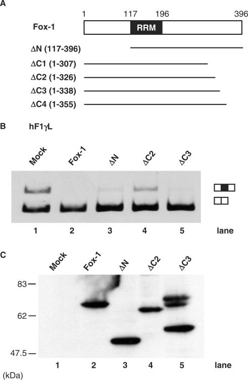
Fox-1 is a regulator of tissue-specific splicing, via binding to the element (U)GCAUG in mRNA precursors, in muscles and neuronal cells. Fox-1 can regulate splicing positively or negatively, most likely depending on where it binds relative to the regulated exon. In cases where the (U)GCAUG element lies in an intron upstream of the alternative exon, Fox-1 protein functions as a splicing repressor to induce exon skipping. Here we report the mechanism of exon skipping regulated by Fox-1, using the hF1gamma gene as a model system. We found that Fox-1 induces exon 9 skipping by repressing splicing of the downstream intron 9 via binding to the GCAUG repressor elements located in the upstream intron 8. In vitro splicing analyses showed that Fox-1 prevents formation of the pre-spliceosomal early (E) complex on intron 9. In addition, we located a region of the Fox-1 protein that is required for inducing exon skipping. Taken together, our data show a novel mechanism of how RNA-binding proteins regulate alternative splicing.


Figures

Figure 1.
Figure 1.
Mouse Fox-1 induces exon 9 skipping of hF1γ pre-mRNA via binding to GCAUG element. (A) The schematic representation of various hF1γ mini-genes. The Fox-1-binding sequence GCAUG and its mutated sequence CGAUG are shown as open and closed circles, respectively. (B) Transfection assays of various hF1γ mini-genes into CV-1 cells. The hF1γL (lanes 1–3), hF1γS (lanes 4–6), hF1γSmt (lanes 7–9) and hF1γSmt+3GCAUG mini-genes (lanes 10–12) were co-expressed with pCS2+MT vector (lanes 1, 4, 7, 10), Fox-1 (lanes 2, 5, 8, 11) and F-A mutant (lanes 3, 6, 9, 12). Splicing products were analyzed by RT-PCR. Splicing products are schematically shown on the right. All experiments were performed more than three times. Average percentage and SD of exon 9 exclusion are shown at the bottom of each lane. (C) Upper panel shows western blotting of cell extracts to detect Fox-1 proteins: Mock, Fox-1 and Fox-1 F-A, expressed from the pCS2+MT vector using the anti-Myc antibody. The positions of molecular size markers are shown on the right. Lower panel shows western blotting of the same cell extracts with anti-U2AF antibody as a loading control.
Figure 2.
Figure 2.
Mouse Fox-1 induces exon 9 skipping of hF1γ pre-mRNA by repressing the splicing of intron 9 via binding to GCAUG element in intron 8. (A) Analysis of intron 8 splicing in CV-1 cells. The Ex8-9 (lanes 1–3) and Ex8-9 mt mini-genes (lanes 4–6) were co-transfected with pCS2+MT vector (lanes 1 and 4), mFox-1 (lanes 2 and 5), F-A (lanes 3 and 6). The splicing products were analyzed by RT-PCR. A GFP plasmid was cotransfected as an internal reference for transfection efficiency, RNA recovery and loading. The positions of spliced products and GFP are indicated on the right. Average percentage and SD of splicing efficiency are shown at the bottom of each lane. (B) Analysis of intron 9 splicing in CV-1 cells. Transfection analyses of the Ex9-10 (lanes 1–3) and Ex9-10 mt mini-genes (lanes 4–6) with pCS2+MT vector (lanes 1 and 4), mFox-1 (lanes 2 and 5), F-A (lanes 3 and 6). The positions of spliced products and GFP are indicated on the right. Average percentage and SD of splicing efficiency are shown at the bottom of each lane. (C) Transfection analyses of the BPmt (lanes 1–3) and 5′SSmt (lanes 4–6) mini-genes with pCS2+MT vector (lanes 1 and 4), mFox-1 (lanes 2 and 5), F-A (lanes 3 and 6). Schematic representation of mini-genes is shown on the left of each panel. Open and closed circles show the GCAUG element and its mutated element, CGAUG, respectively. Open and closed triangles show branch point (BP) and its mutated site, respectively. A cross represents a mutated 5′ splice site in intron 9. Sequences of these elements are shown at the bottom. A bold letter represents the branch point nucleotide.
Figure 3.
Figure 3.
Fox-1 represses the intron 9 splicing in vitro. (A) Western blotting of the nuclear extracts using anti-Flag and anti-U2AF antibodies. HeLa cell nuclear extracts were mixed with nuclear extracts of HEK293 cells transfected with pCS2-Flag vector alone (Mock) or pCS2 Flag-mFox-1 (Flag Fox-1). The positions of molecular size markers are shown on the right side of upper panel. In addition to the band at the expected size, an additional band was detected (asterisk). (B) In vitro splicing reaction of Ex9-10 + 3GCAUG (lanes 1–4) and Ex9-10ΔGCAUG (lanes 5–8) in Mock nuclear extracts (lanes 2, 3, 7 and 8) or nuclear extracts containing Flag-tagged mFox-1 (lanes 4, 5, 9 and 10) for indicated time above each lane. Pre-mRNAs and splicing products are indicated schematically on the right.
Figure 4.
Figure 4.
Fox-1 blocks formation of the pre-spliceosomal early (E) complex on intron 9 via binding to GCAUG element in intron 8. (A) Spliceosomal complex formation on the Ex9-10 + 3GCAUG (lanes 1–4) and the Ex9-10 ΔGCAUG (lanes 1, 2, 5 and 6) transcripts in Mock nuclear extracts (lanes 1 and 2) or nuclear extracts containing Flag-tagged mFox-1 (lanes 3, 4, 7 and 8) under normal splicing condition in the presence of ATP for the indicated time above each lane. Spliceosomal complexes were separated by 4% native polyacrylamide gels and position of each complex is indicated on the left. (B) Spliceosomal complex formation on the hF1γEx 9-10 transcripts in the absence of ATP. Transcripts of the Ex9-10 + 3GCAUG (lanes 1–4) and the Ex9-10 ΔGCAUG (lanes 5–8) were incubated in Mock nuclear extracts (lanes 1, 2, 5 and 6) or Fox-1 nuclear extracts (lanes 3, 4, 7 and 8) in the absence of ATP and separated on a 1.5% native agarose gel. Positions of pre-spliceosomal complexes E and H are indicated on the left.
Figure 5.
Figure 5.
Identification of the functional region of Fox-1 protein required for alternative exon skipping. (A) Diagrams of full-length and truncated mutants of mFox-1 proteins. (B) Transfection analyses of the hF1γ L mini-gene. The hF1γ L mini-gene (lanes 1–5) were co-expressed in CV-1 cells along with pCS2+MT vector (lane 1), mFox-1 (lane 2), ΔN (lane 3), ΔC2 (lane 4), and ΔC3 (lane 5). The splicing products were analyzed by RT-PCR. The positions of spliced products are indicated on the right. (C) Western blotting of cell extracts to detect Fox-1 truncated mutants of Fox-1 proteins: Each of the truncated mutants was expressed from the pCS2+MT vector using the anti-Myc antibody. In lane 5, additional slow migrating bands as well as an expected band for ΔC3 protein were observed.

Similar articles

Cited by

References

    1. Graveley BR. Alternative splicing: increasing diversity in the proteomic world. Trends Genet. 2001;17:100–107. - PubMed
    1. Maniatis T, Tasic B. Alternative pre-mRNA splicing and proteome expansion in metazoans. Nature. 2002;418:236–243. - PubMed
    1. Black DL. Mechanisms of alternative pre-messenger RNA splicing. Annu. Rev. Biochem. 2003;72:291–336. - PubMed
    1. Das R, Reed R. Resolution of the mammalian E complex and the ATP-dependent spliceosomal complexes on native agarose mini-gels. RNA. 1999;5:1504–1508. - PMC - PubMed
    1. Kent OA, Ritchie DB, Macmillan AM. Characterization of a U2AF-independent commitment complex (E′) in the mammalian spliceosome assembly pathway. Mol. Cell. Biol. 2005;25:233–240. - PMC - PubMed

Publication types

MeSH terms