. 2007 Oct;81(20):11468-78.
doi: 10.1128/JVI.01364-07. Epub 2007 Aug 8.

Intracellular trafficking and maturation of herpes simplex virus type 1 gB and virus egress require functional biogenesis of multivesicular bodies

Affiliations

Intracellular trafficking and maturation of herpes simplex virus type 1 gB and virus egress require functional biogenesis of multivesicular bodies

Arianna Calistri et al. J Virol. 2007 Oct.

Abstract

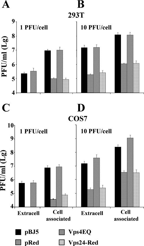
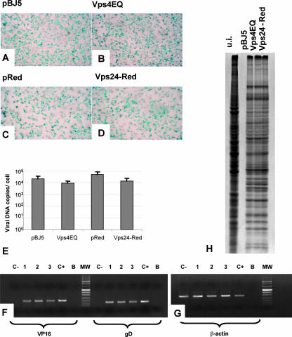
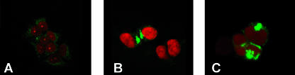
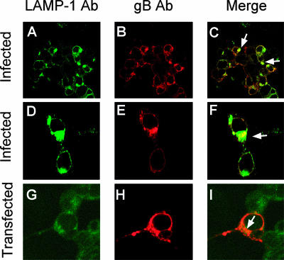
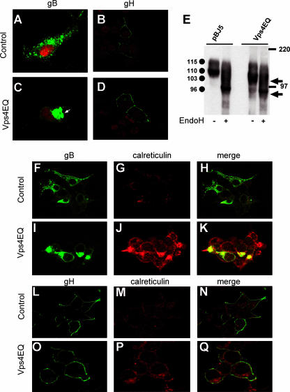
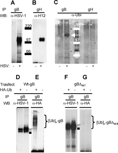
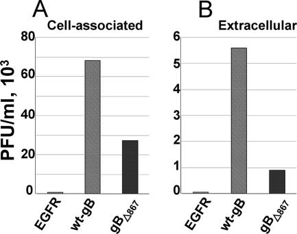
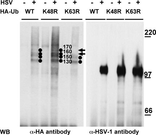
The biogenesis of multivesicular bodies (MVBs) is topologically equivalent to virion budding. Hence, a number of viruses exploit the MVB pathway to build their envelope and exit from the cell. By expression of dominant negative forms of Vps4 and Vps24, two components of the MVB pathway, we observed an impairment in infectious herpes simplex virus (HSV) assembly/egress, in agreement with a recent report showing the involvement in HSV envelopment of Vps4, the MVB-specific ATPase (C. M. Crump, C. Yates, and T. Minson, J. Virol. 81:7380-7387). Furthermore, HSV infection resulted in morphological changes to MVBs. Glycoprotein B (gB), one of the most highly conserved glycoproteins across the Herpesviridae family, was sorted to MVB membranes. In cells expressing the dominant negative form of Vps4, the site of intracellular gB accumulation was altered; part of gB accumulated as an endoglycosidase H-sensitive immature form at a calreticulin-positive compartment, indicating that gB traffic was dependent on a functional MVB pathway. gB was ubiquitinated in both infected and transfected cells. Ubiquitination was in part dependent on ubiquitin lysine 63, a signal for cargo sorting to MVBs. Partial deletion of the gB cytoplasmic tail resulted in a dramatic reduction of ubiquitination, as well as of progeny virus assembly and release to the extracellular compartment. Thus, HSV envelopment/egress and gB intracellular trafficking are dependent on functional MVB biogenesis. Our data support the view that the sorting of gB to MVB membranes may represent a critical step in HSV envelopment and egress and that modified MVB membranes constitute a platform for HSV cytoplasmic envelopment or that MVB components are recruited to the site(s) of envelopment.


Figures

FIG. 1.
FIG. 1.
Effect of Vps4E228Q and Vps24-Red on the yield of extracellular and cell-associated HSV-1. 293T cells (A, B) and COS-7 cells (C, D) were transfected either with an empty vector (pBJ5 or pDsRed2-N1) or with constructs expressing either Vps4E228Q or Vps24-Red. Twelve hours after transfection, the cells were infected with HSV-1 at 1 PFU/cell (A, C) or 10 PFU/cell (B, D). At 36 h postinfection, the extracellular (Extracell) and cell-associated virus were titrated by plaque assay in Vero cells.
FIG. 2.
FIG. 2.
Vps4E228Q (Vps4EQ) or Vps24-Red does not affect the early steps of HSV-1 replication. (A to D) 293T cells were transfected with plasmids expressing the dominant negative forms of Vps4 (B) or Vps24 (D) cellular protein or their respective empty vector, pBJ5 (A) or pDsRed2-N1 (pReD) (C). Twelve hours after transfection, the cells were infected with the R8102 recombinant HSV-1, carrying the lacZ gene under the control of the ICP-27 immediate promoter (3 PFU/cell) (5). Six hours later, the infected cells were stained with X-Gal. The panels illustrate light microscope images (magnification, ×1.5). (E) Quantification of HSV-1 genomic copies by real-time PCR assay. 293T cells were transfected with the constructs expressing either Vps4E228Q or Vps24-Red or the corresponding empty vectors. Twelve hours posttransfection, the cells were infected with HSV-1 (10 PFU/cell). Twenty-four hours later, the amount of viral DNA was evaluated by real-time PCR assay. Results are expressed as numbers of viral DNA copies per cell. (F and G) Total RNA was extracted from 293T cells transfected with the indicated plasmids. The RT-PCR assay was performed with primers specific for VP16 and gD (F) or the β-actin genes (G). Lanes: 1, 293T cells transfected with pBJ5 and infected with HSV-1; 2, 293T cells transfected with pBJ5-Vps4E228Q and infected with HSV-1; 3, 293T cells transfected with pDsRed-Vps24 and infected with HSV-1; C+, Vero cells infected with HSV-1; C−, uninfected 293T cells; B, no-template control; MW, molecular weight markers. (H) 293T cells transfected with the indicated plasmids were infected with HSV-1 strain F, labeled with [35S]methionine, and harvested 24 h after infection. Radiolabeled proteins were detected by autoradiography following SDS-PAGE. u.i., uninfected 293T cells.
FIG. 3.
FIG. 3.
Effect of HSV infection on the morphology of the MVB compartment. 293T cells were infected with HSV-1 and analyzed at 12 or 24 h after infection by immunofluorescence with PAb to LAMP-1. The cell nuclei were stained with propidium iodide. Cells were observed with a Leica confocal microscope at a ×63 magnification objective. (A) Uninfected 293T cells. (B) 293T cells 12 h after HSV-1 infection. (C) 293T cells 24 h after HSV-1 infection.
FIG. 4.
FIG. 4.
gB accumulates in part at MVB membranes in HSV-infected and -transfected cells. (A to F) 293T cells were infected with HSV-1 (10 PFU/cell). Twenty-four hours later, the cells were stained with MAb to gB (Virusys) and PAb to LAMP-1 and analyzed by confocal microscopy at a ×63 magnification objective. (G to I) 293T cells were transfected with a construct expressing gB (pgBwt-MTS). Fourty-eight hours later, the cells were stained with MAb to gB (Virusys) and a PAb to LAMP-1 and analyzed by confocal microscopy. The arrows point to colocalization spots. (A to C) Low magnification; (D to F) high magnification.
FIG. 5.
FIG. 5.
Effect of blocking MVB biogenesis on gB and gH localization and maturation. 293T cells were cotransfected with a construct expressing Vps4E228Q (Vps4EQ) or the empty vector pBJ5, along with the pgBwt-MTS plasmid, expressing wt gB (A and C), or gH-MTS-gL-MTS plasmids encoding gH/gL (B and D). At 48 h after transfection, the cells were stained with MAb H1817 to gB (A and C) or MAb 53S to gH (B, D) and analyzed by confocal microscopy. Nuclei were stained with propidium iodide. (E) 293T cells were cotransfected with a construct expressing Vps4E228Q or the empty vector pBJ5, along with the pgBwt-MTS plasmid. Forty-eight hours after transfection, gB was immunoprecipitated from the cell lysates and treated with endo H (+) or left undigested (−). Samples were analyzed by SDS-PAGE and Western blotting with PAb to the major HSV-1 glycoproteins. Circles identify the gB forms exhibiting the indicated apparent Mrs. Arrows identify the electrophoretic mobilities of endo H-digested immature forms of gB. The electrophoretic mobilities of the molecular mass markers are reported. (F to Q) 293T cells cotransfected with pBJ5-Vps4E228Q along with either pgBwt-MTS (I to K) or gH-MTS-gL-MTS (O to Q) were analyzed by confocal microscopy, with MAb to gB (Virusys) or MAb 53S to gH and a PAb to calreticulin, as indicated. 293T cells cotransfected with the empty vector pBJ5 together with either the pgBwt-MTS plasmid (F to H) or the gH-MTS-gL-MTS plasmids (L to N) were used as a control.
FIG. 6.
FIG. 6.
HSV-1 gB is ubiquitinated in infected and in transfected cells. (A to C) Electrophoretic and Western blotting analyses of gB and gH immunoprecipitated (IP) from lysates of HSV-1-infected (+) or uninfected (−) 293T cells by means of MAb H1817 to gB or MAb 53S to gH. The immunoprecipitated proteins were separated by SDS-PAGE, followed by Western blotting (WB) with the indicated antibodies (α-HSV-1, PAb to the major HSV-1 glycoproteins; α-H12, MAb to gH antibody; α-Ubi, PAb to ubiquitin). (C) The arrow points to the band of ubiquitinated gB. Numbers identify the electrophoretic mobilities of the molecular mass markers. (D to G) 293T cells were cotransfected with a construct expressing wt gB (pgBwt-MTS) (D, E) or a truncated form of gB (gBΔ867) (F, G). Cells were also transfected with a construct encoding HA-tagged wt ubiquitin (HA-Ub) or the corresponding pBJ5 empty vector, indicated as HA-Ub + or HA-Ub −, respectively. Forty-eight hours posttransfection, gB was immunoprecipitated with MAb H1817. The separated proteins were analyzed by Western blotting with the antibodies listed above. Braces identify polyubiquitinated forms of gB. 97 indicates the electrophoretic mobility of the molecular mass marker.
FIG. 7.
FIG. 7.
Yield of cell-associated and extracellular virions complemented with gBΔ867, wt gB, or EGFR. The ΔgB HSV KΔt (9) was grown in 293T cells transiently expressing wt gB, gBΔ867, or EGFR as a negative control. At 24 h after infection, the cell-associated progeny virus (A) or virus released in the extracellular medium (B) was titrated in gB-complementing D6 cells.
FIG. 8.
FIG. 8.
Effect of ubiquitin mutants on gB ubiquitination. To detect ubiquitin conjugates, 293T cells were transfected with expression vectors encoding the following HA-tagged forms of ubiquitin (HA-Ub): the wt, K48R mutant, or K63R mutant. Twenty-four hours later, the cells were infected with HSV-1 (10 PFU/cell) (+) or mock infected (−). Twenty-four hours postinfection, gB was immunoprecipitated from cell lysates. Western blotting (WB) was performed with the indicated antibodies (α-HA, MAb to HA; α-HSV-1, PAb to major HSV-1 glycoproteins). Circles identify the ubiquitinated forms of gB; values to the right of the circles indicate their apparent Mrs. Arrows point to the 170- and 160-kDa forms of ubiquitinated gB absent from the K63R sample. The values to the right identify the migration positions of molecular mass markers (in kilodaltons).

References

    1. Avitabile, E., S. Di Gaeta, M. R. Torrisi, P. L. Ward, B. Roizman, and G. Campadelli-Fiume. 1995. Redistribution of microtubules and Golgi apparatus in herpes simplex virus-infected cells and their role in viral exocytosis. J. Virol. 69:7472-7482. - PMC - PubMed
    1. Avitabile, E., G. Lombardi, T. Gianni, M. Capri, and G. Campadelli-Fiume. 2004. Coexpression of UL20p and gK inhibits cell-cell fusion mediated by herpes simplex virus glycoproteins gD, gH-gL, and wild-type gB or an endocytosis-defective gB mutant and downmodulates their cell surface expression. J. Virol. 78:8015-8080. - PMC - PubMed
    1. Babst, M. 2005. A protein's final ESCRT. Traffic 6:2-9. - PubMed
    1. Baghian, A., L. Huang, S. Newman, S. Jayachandra, and K. G. Kousoulas. 1993. Truncation of the carboxy-terminal 28 amino acids of glycoprotein B specified by herpes simplex virus type 1 mutant amb1511-7 causes extensive cell fusion. J. Virol. 67:2396-2401. - PMC - PubMed
    1. Baines, J. D., and B. Roizman. 1991. The open reading frames UL3, UL4, UL10, and UL16 are dispensable for the replication of herpes simplex virus 1 in cell culture. J. Virol. 65:938-944. - PMC - PubMed

Publication types

Substances