. 2007 Oct;81(20):11267-81.
doi: 10.1128/JVI.00007-07. Epub 2007 Aug 8.

Antisense transcription in the human cytomegalovirus transcriptome

Affiliations

Antisense transcription in the human cytomegalovirus transcriptome

Guojuan Zhang et al. J Virol. 2007 Oct.

Abstract

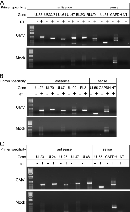
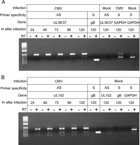
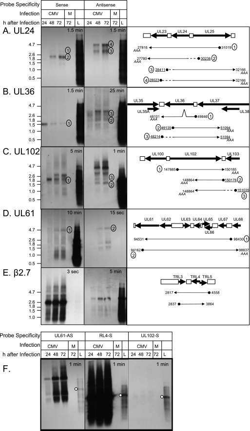
Human cytomegalovirus (HCMV) infections are prevalent in human populations and can cause serious diseases, especially in those with compromised or immature immune systems. The HCMV genome of 230 kb is among the largest of the herpesvirus genomes. Although the entire sequence of the laboratory-adapted AD169 strain of HCMV has been available for 18 years, the precise number of viral genes is still in question. We undertook an analysis of the HCMV transcriptome as an approach to enumerate and analyze the gene products of HCMV. Transcripts of HCMV-infected fibroblasts were isolated at different times after infection and used to generate cDNA libraries representing different temporal classes of viral genes. cDNA clones harboring viral sequences were selected and subjected to sequence analysis. Of the 604 clones analyzed, 45% were derived from genomic regions predicted to be noncoding. Additionally, at least 55% of the cDNA clones in this study were completely or partially antisense to known or predicted HCMV genes. The remarkable accumulation of antisense transcripts during infection suggests that currently available genomic maps based on open-reading-frame and other in silico analyses may drastically underestimate the true complexity of viral gene products. These findings also raise the possibility that aspects of both the HCMV life cycle and genome organization are influenced by antisense transcription. Correspondingly, virus-derived noncoding and antisense transcripts may shed light on HCMV pathogenesis and may represent a new class of targets for antiviral therapies.


Figures

FIG. 1.
FIG. 1.
Verification of virally derived AS transcripts. Digital images of agarose gels used to separate PCR products specific for virally derived AS and S transcripts. Confluent MRC-5 cells in six-well tissue culture plates were exposed to 2 PFU per cell of the AD169 strain of HCMV (top panels) or were mock infected (bottom panels). (A) RT-PCR of AS transcripts identified in the E library using total RNA isolated at 48 h after infection. (B) RT-PCR of AS transcripts identified in the L library using total RNA isolated at 72 h after infection. (C) RT-PCR of AS transcripts specific to tegument genes using total RNA isolated at 24 h after infection (UL47) or 72 h after infection (UL23, UL24, UL25, and UL85). Total RNA was isolated and subjected to RT-PCR using primers specific to virally derived AS transcripts (Table 1). Primers specific for the S transcripts of viral UL55 (gB) or cellular GAPDH were included as positive controls. No-reverse-transcriptase and no-template (NT) controls were run in parallel. At 48 h, the no-template control included primers specific for AS US30/31. At 72 h, the no-template control included primers specific for AS UL87/88. In panel C, the no-template control included primers specific for AS UL23. RT-PCR products were separated by agarose gel (1% [wt/vol]) electrophoresis, and bands were visualized by exposure to UV light.
FIG. 2.
FIG. 2.
Verification of AS transcripts in endothelial cells infected with a clinical strain of HCMV. Digital image of an agarose gel used to separate PCR products specific for virally derived AS and S transcripts. Confluent HUVEC monolayers in six-well tissue culture plates were inoculated with the VHL/E strain of HCMV (1 PFU/cell). Cells were harvested at 24, 48, 72, 96, and 120 h postinfection or were mock infected for 120 h. Total RNA was isolated and subjected to RT-PCR using primers specific to virally derived AS transcripts for UL36/37 (A) or UL102 (B). Primers specific for the S transcripts of viral UL55 (gB) or cellular GAPDH were included as positive controls. No-reverse-transcriptase and no-template controls were run in parallel. RT-PCR products were separated by agarose gel (1% [wt/vol]) electrophoresis, and bands were visualized by exposure to UV light.
FIG. 3.
FIG. 3.
Northern blot analysis of S and AS transcripts. Film images of S and AS transcripts analyzed by Northern blotting and schematic depictions of select S and AS transcripts. Confluent MRC-5 cells in six-well tissue culture plates were exposed to 2 PFU per cell of the AD169 strain of HCMV or were mock infected (M). Cells were harvested at 24, 48, and 72 h after infection. Total RNA was isolated, subjected to denaturing agarose gel electrophoresis, and transferred to nylon membranes. Prelabeled RNA molecular mass markers were loaded for each group (L). Membranes were incubated with probes specific for the S and AS transcripts of UL24, 3 μg RNA/lane (A); UL36, 3 μg RNA/lane (B); UL102, 4 μg RNA/lane (C and F); UL61, 5 μg RNA/lane (D and F); or RL4, 4 μg RNA/lane (E and F) as described in Materials and Methods. Exposure times are indicated at the top of each panel. A schematic of transcripts represented by select cDNA clones isolated in our library relative to the genome is shown in the right panels. Gene regions and intergenic regions are depicted by thick arrows and white boxes, respectively. Transcripts cloned in this study are represented as thin arrows below the gene regions. 5′ ends of transcripts are depicted with filled circles. Clones in which we identified the poly(A) tails are indicated (AAA). The genomic positions of the 5′ and 3′ ends of the clones in the libraries are shown. Underlined genomic positions are those verified by RACE. Dashed lines represent presumptive transcript sequences based on RACE analysis. Tentative assignment of bands corresponding to clones identified in this study is indicated with circled numbers. In panel F, the relative abundances of transcripts overlapping the RL4 and UL61 gene regions are compared to those of transcripts derived from the UL102 gene region. The white circle indicates the position of the 2.6-kb marker band. No images were altered.

Similar articles

Cited by

References

    1. Alderete, J. P., S. J. Child, and A. P. Geballe. 2001. Abundant early expression of gpUL4 from a human cytomegalovirus mutant lacking a repressive upstream open reading frame. J. Virol. 75:7188-7192. - PMC - PubMed
    1. Atalay, R., A. Zimmermann, M. Wagner, E. Borst, C. Benz, M. Messerle, and H. Hengel. 2002. Identification and expression of human cytomegalovirus transcription units coding for two distinct Fcγ receptor homologs. J. Virol. 76:8596-8608. - PMC - PubMed
    1. Awasthi, S., J. A. Isler, and J. C. Alwine.2004. Analysis of splice variants of the immediate-early 1 region of human cytomegalovirus. J. Virol. 78:8191-8200. - PMC - PubMed
    1. Baldick, C. J., Jr., and T. Shenk. 1996. Proteins associated with purified human cytomegalovirus particles. J. Virol. 70:6097-6105. - PMC - PubMed
    1. Bankier, A. T., S. Beck, R. Bohni, C. M. Brown, R. Cerny, M. S. Chee, C. A. Hutchison III, T. Kouzarides, J. A. Martignetti, E. Preddie, et al. 1991. The DNA sequence of the human cytomegalovirus genome. DNA Seq. 2:1-12. - PubMed

Publication types

LinkOut - more resources