

17beta-Estradiol attenuates diabetic kidney disease by regulating extracellular matrix and transforming growth factor-beta protein expression and signaling
- PMID: 17686959
- PMCID: PMC3179625
- DOI: 10.1152/ajprenal.00079.2007
17beta-Estradiol attenuates diabetic kidney disease by regulating extracellular matrix and transforming growth factor-beta protein expression and signaling
Abstract
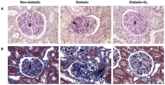
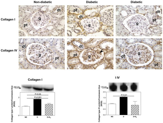
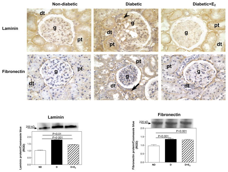
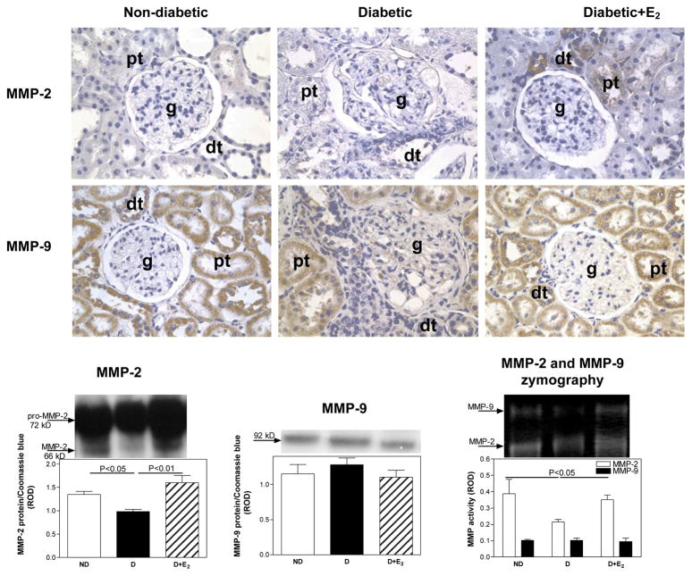
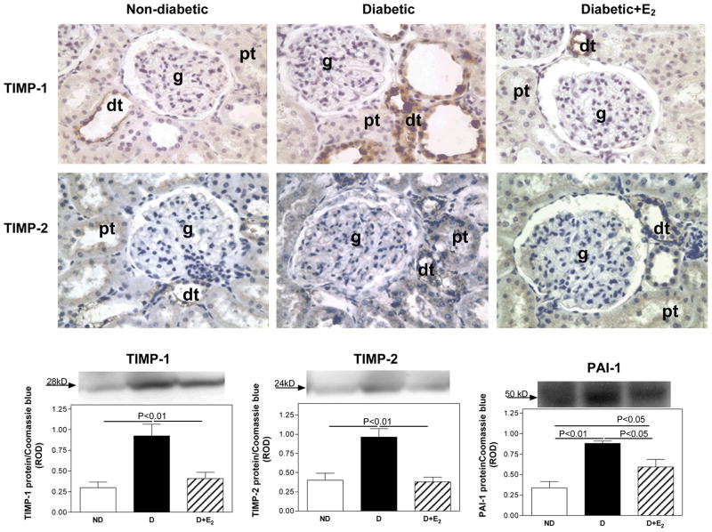
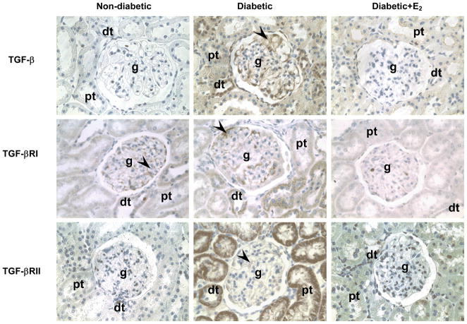
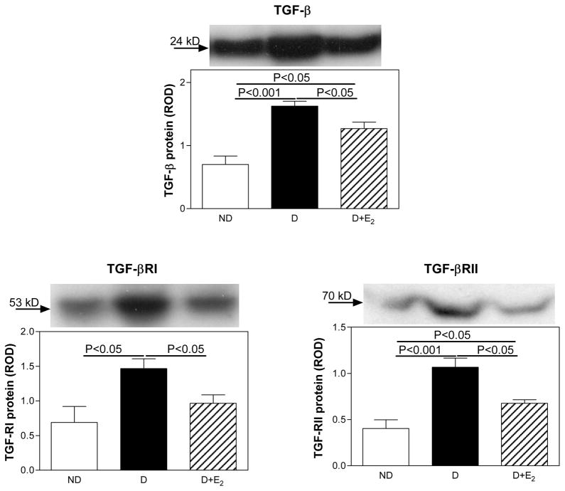
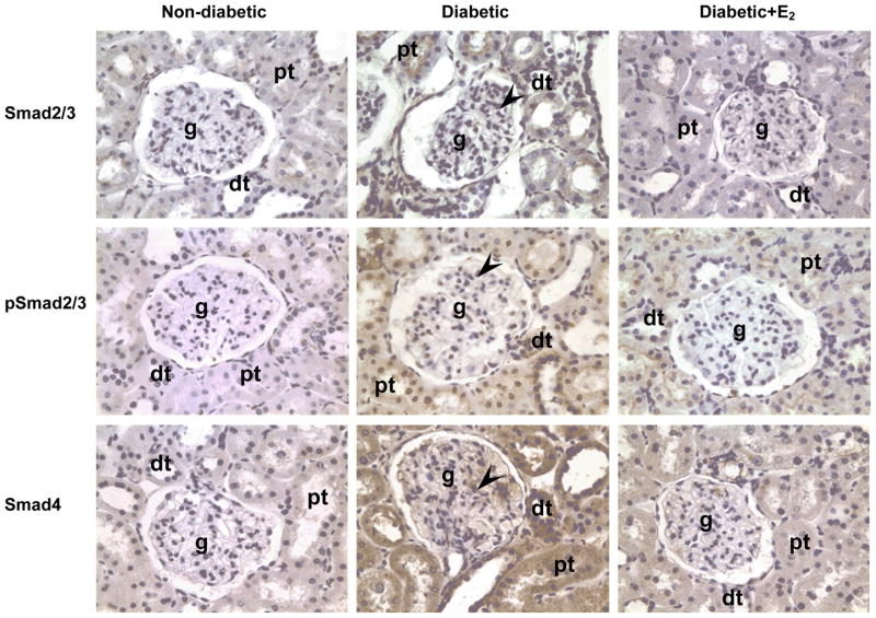
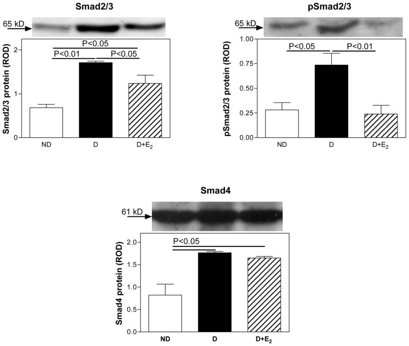
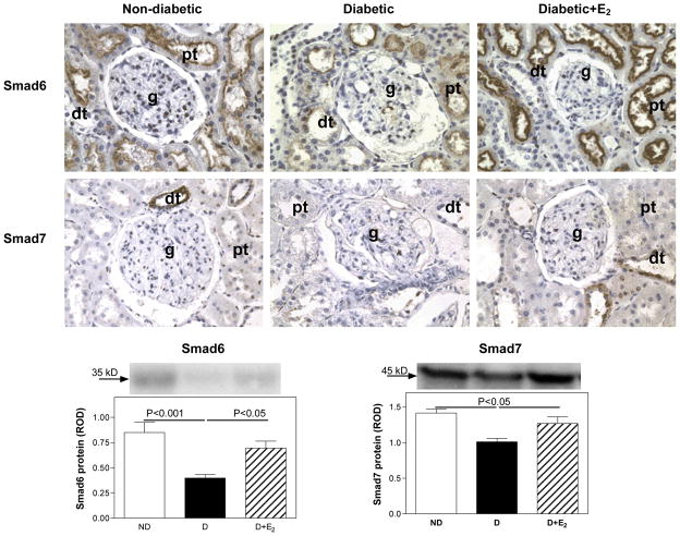
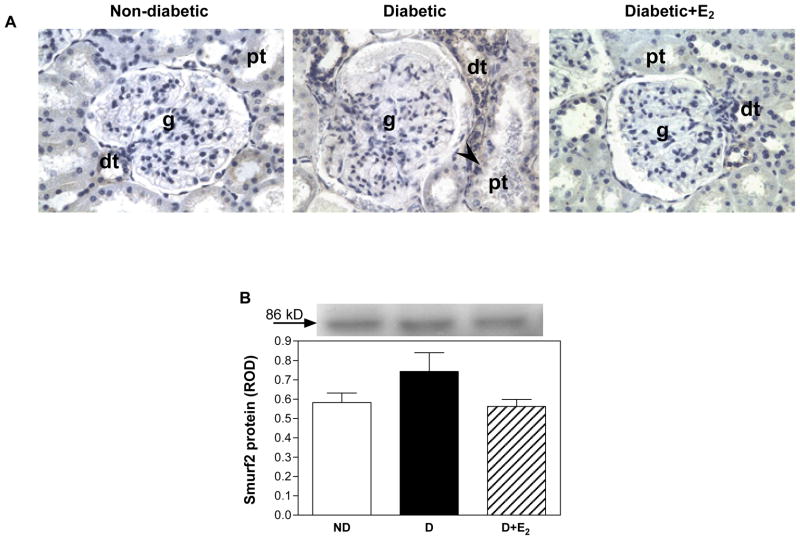
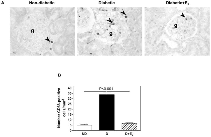
We previously showed that supplementation with 17beta-estradiol (E2) from the onset of diabetes attenuates the development of diabetic renal disease. The aim of the present study was to examine whether E2 can also attenuate the disease process once it has developed. The present study was performed in nondiabetic and streptozotocin-induced diabetic Sprague-Dawley rats. E2 supplementation began after 9 wk of diabetes and continued for 8 wk. Diabetes was associated with an increase in urine albumin excretion, glomerulosclerosis, tubulointerstitial fibrosis, renal cortical collagen type I and IV, laminin, plasminogen activator inhibitor-1, tissue inhibitors of metalloproteinase-1 and -2, transforming growth factor (TGF)-beta, TGF-beta receptor type I and II, Smad2/3, phosphorylated Smad2/3, and Smad4 protein expression, and CD68-positive cell abundance. Decreases in matrix metalloproteinase (MMP)-2 protein expression and activity and decreases in Smad6 and Smad7 protein expression were also associated with diabetes. E2 supplementation completely or partially attenuated all these changes, except Smad4 and fibronectin, on which E2 supplementation had no effect. These data suggest that E2 attenuates the progression of diabetic renal disease once it has developed by regulating extracellular matrix, TGF-beta, and expression of its downstream regulatory proteins. These findings support the notion that sex hormones in general, and E2 in particular, are important regulators of renal function and may be novel targets for the treatment and prevention of diabetic renal disease.
Figures
Similar articles
-
17beta-Estradiol supplementation reduces tubulointerstitial fibrosis by increasing MMP activity in the diabetic kidney.Am J Physiol Regul Integr Comp Physiol. 2007 Feb;292(2):R769-77. doi: 10.1152/ajpregu.00375.2006. Epub 2006 Aug 24. Am J Physiol Regul Integr Comp Physiol. 2007. PMID: 16931652
-
Imbalance in sex hormone levels exacerbates diabetic renal disease.Hypertension. 2008 Apr;51(4):1218-24. doi: 10.1161/HYPERTENSIONAHA.107.100594. Epub 2008 Feb 7. Hypertension. 2008. PMID: 18259041 Free PMC article.
-
Diabetic nephropathy and transforming growth factor-beta: transforming our view of glomerulosclerosis and fibrosis build-up.Semin Nephrol. 2003 Nov;23(6):532-43. doi: 10.1053/s0270-9295(03)00132-3. Semin Nephrol. 2003. PMID: 14631561 Review.
-
Fibroblast Growth Factor 21 Attenuates Diabetes-Induced Renal Fibrosis by Negatively Regulating TGF-β-p53-Smad2/3-Mediated Epithelial-to-Mesenchymal Transition via Activation of AKT.Diabetes Metab J. 2020 Feb;44(1):158-172. doi: 10.4093/dmj.2018.0235. Epub 2019 Oct 28. Diabetes Metab J. 2020. PMID: 31701691 Free PMC article.
-
Estrogens and the diabetic kidney.Gend Med. 2008;5 Suppl A(Suppl A):S103-13. doi: 10.1016/j.genm.2008.03.010. Gend Med. 2008. PMID: 18395675 Free PMC article. Review.
Cited by
-
Proteases in Plasma and Kidney of db/db Mice as Markers of Diabetes-Induced Nephropathy.ISRN Endocrinol. 2011;2011:832642. doi: 10.5402/2011/832642. Epub 2011 Aug 4. ISRN Endocrinol. 2011. PMID: 22363890 Free PMC article.
-
(Pro)renin receptor contributes to renal mitochondria dysfunction, apoptosis and fibrosis in diabetic mice.Sci Rep. 2019 Aug 12;9(1):11667. doi: 10.1038/s41598-019-47055-1. Sci Rep. 2019. PMID: 31406124 Free PMC article.
-
Myocardin-Related Transcription Factor A Epigenetically Regulates Renal Fibrosis in Diabetic Nephropathy.J Am Soc Nephrol. 2015 Jul;26(7):1648-60. doi: 10.1681/ASN.2014070678. Epub 2014 Oct 27. J Am Soc Nephrol. 2015. PMID: 25349198 Free PMC article.
-
Renoprotective effects of estrogen on acute kidney injury: the role of SIRT1.Int Urol Nephrol. 2021 Nov;53(11):2299-2310. doi: 10.1007/s11255-020-02761-y. Epub 2021 Jan 17. Int Urol Nephrol. 2021. PMID: 33458788 Review.
-
Unraveling Sex Differences in Kidney Health and CKD: A Review of the Effect of Sex Hormones.Clin J Am Soc Nephrol. 2024 Dec 13;20(2):301-10. doi: 10.2215/CJN.0000000642. Online ahead of print. Clin J Am Soc Nephrol. 2024. PMID: 39671256
References
-
- Animal models of diabetic complications Consortium. Validation of mouse models of diabetic nephropathy. http://wwwamdccorg/
-
- Antus B, Hamar P, Kokeny G, Szollosi Z, Mucsi I, Nemes Z, Rosivall L. Estradiol is nephroprotective in the rat remnant kidney. Nephrol Dial Transplant. 2003;18:54–61. - PubMed
-
- Benigni A, Zoja C, Campana M, Corna D, Sangalli F, Rottoli D, Gagliardini E, Conti S, Ledbetter S, Remuzzi G. Beneficial effect of TGFbeta antagonism in treating diabetic nephropathy depends on when treatment is started. Nephron Exp Nephrol. 2006;104:158–168. - PubMed
-
- Birch Nielsen C, Krag S, ROS, Flyvbjerg A, Nyengaard J, Forman A, Wogensen L. Transforming growth factor beta1-induced glomerulopathy is prevented by 17beta-estradiol supplementation. Virchows Arch. 2004;444:561–566. - PubMed
Publication types
MeSH terms
Substances
Grants and funding
LinkOut - more resources
Full Text Sources
Medical
Miscellaneous