. 2007 Aug 14;104(33):13355-60.
doi: 10.1073/pnas.0706121104. Epub 2007 Aug 8.

Expression of dominant-negative Dmp53 in the adult fly brain inhibits insulin signaling

Affiliations

Expression of dominant-negative Dmp53 in the adult fly brain inhibits insulin signaling

Johannes H Bauer et al. Proc Natl Acad Sci U S A. .

Abstract

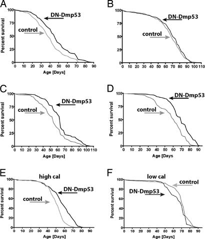
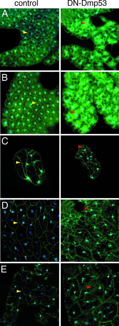
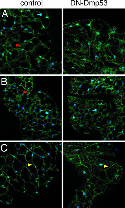
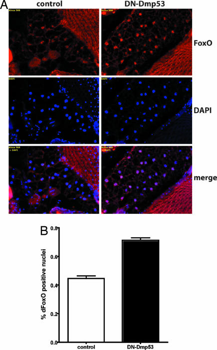
In Drosophila melanogaster, p53 (Dmp53) is an important mediator of longevity. Expression of dominant-negative (DN) forms of Dmp53 in adult neurons, but not in muscle or fat body cells, extends lifespan. The lifespan of calorie-restricted flies is not further extended by simultaneously expressing DN-Dmp53 in the nervous system, indicating that a decrease in Dmp53 activity may be a part of the CR lifespan-extending pathway in flies. In this report, we show that selective expression of DN-Dmp53 in only the 14 insulin-producing cells (IPCs) in the brain extends lifespan to the same extent as expression in all neurons and this lifespan extension is not additive with CR. DN-Dmp53-dependent lifespan extension is accompanied by reduction of Drosophila insulin-like peptide 2 (dILP2) mRNA levels and reduced insulin signaling (IIS) in the fat body, which suggests that Dmp53 may affect lifespan by modulating insulin signaling in the fly.


Conflict of interest statement

The authors declare no conflict of interest.

Figures

Fig. 1.
Fig. 1.
Expression of DN-Dmp53 in IPCs extends lifespan and is related to CR. (AD) Survivorship curves of female DN-Dmp53 (black) or isogenic control flies (gray) crossed to various constitutively active neuron-specific drivers are shown. Pan-neuronal (A) (ELAV driver) expression of DN-Dmp53 extends mean lifespan up to 19%. (B) No lifespan extension is seen when a driver specific for dopaminergic neurons is used. When expressed in IPCs [dILP2 -Shen driver (C); dILP2 -Rulifson driver (D)] lifespan extension similar to ELAV driver is observed (18% and 19% extension of mean lifespan for Shen and Rulifson driver, respectively). (E and F) Survivorship curves of dILP2-DN-Dmp53 flies under CR conditions, using the Shen driver. Expression of DN-Dmp53 (black) in IPCs on high calorie food leads to a 14% increase in mean lifespan (E) over controls (gray). When flies are raised on lifespan-extending low calorie food (gray) (F), 37% extension of mean lifespan is observed. Simultaneous expression of DN-Dmp53 (black) does not lead to additional lifespan extension. All experiments shown were performed by using >200 flies each. Mean, median, and maximum lifespan data can be found in Table 1.
Fig. 2.
Fig. 2.
PI3K activity is reduced in long-lived flies expressing DN-Dmp53 in the IPCs or pan-neuronally. The tGPH reporter construct was engineered into the control and the UAS-DN-Dmp53 strain. The resulting lines were then crossed to the dILP2 or ELAV-Switch driver. Fat bodies were isolated from the indicated tissues and analyzed for subcellular localization of the tGPH reporter (blue: DAPI; green: GFP). (A) Fat bodies were isolated from late third-instar of dILP2-DN-Dmp53 larvae. In control flies (Left), plasma membrane staining and no cytoplasic staining is observed. In DN-Dmp53-expressing flies (Right), plasma membrane staining is less pronounced and there is diffuse, cytoplasmic staining. (B) Similar results are observed when fat body cells are isolated from ELAV-Switch-DN-Dmp53 larvae. (CE) When fat body is examined from heads (C) or abdomen (D) adult dILP2-DN-Dmp53 flies, or the abdomen (E) adult ELAV-Switch-DN-Dmp53 flies, control flies show plasma membrane staining with little cytoplasmic staining, whereas there is diffuse cytoplasmic staining in DN-Dmp53 expressing flies. Yellow arrows depict tGPH at the plasma membrane, red arrows cytosolic tGPH. Shown are representative images of at least five independent experiments using three to five flies per experiment.
Fig. 3.
Fig. 3.
The fat body cells of DN-Dmp53-expressing flies still respond to a starvation/sugar refeeding challenge. (A) Adult abdominal fat body from dILP2-DN-Dmp53 flies starved for 5 h shows a general decrease in membrane-associated tGPH in control (Left) and DN-Dmp53-expressing flies (Right) that is greater in DN-Dmp53 expressing flies. (B) After 20 h of starvation, membrane-associated tGPH is completely lost in both control (Left) and DN-Dmp53- expressing flies. (Right) (C) Three-hour feeding of a 5% sucrose solution after a 5-h period of water only starvation leads to almost exclusive plasma membrane staining in both control flies (Left) and flies expressing DN-Dmp53 in the IPCs. (Right) Yellow arrows depict tGPH bound at the plasma membrane, red arrows cytosolic tGPH, and blue arrows show starvation induced cytosolic vacuoles. Shown are representative images of at least five independent experiments using three to five flies each.
Fig. 4.
Fig. 4.
dFoxO accumulates in the nucleus of fat body cells in long-lived dILP2 driven DN-Dmp53 expressing flies. (A) Heads of 10-day-old dILP2-DN-Dmp53 flies were cryosectioned and stained with an α-dFoxO antibody. In control flies (Left), only a small percentage of fat body cells show dFoxO staining. In contrast, in DN-Dmp53 expressing flies (Right) almost all fat body nuclei are positive for dFoxO antibody staining (red, α-dFoxO; blue, DAPI; purple, merge). (B) Quantification of dFoxO nuclear translocation in fat body cells. A total of ≈1,300 individual nuclei were scored for dFoxO staining. Compared with control flies, 30% more nuclei in DN-Dmp53 expressing flies also stain positive for dFoxO. A representative of two independent experiments using at least 10 animals each is shown. Error bars represent the SEM; P < 0.0001.

Similar articles

Cited by

References

    1. Masoro EJ. Sci Aging Knowledge Environ. 2003:RE2. - PubMed
    1. Tatar M, Bartke A, Antebi A. Science. 2003;299:1346–1351. - PubMed
    1. Goberdhan DC, Wilson C. Differentiation. 2003;71:375–397. - PubMed
    1. Obsil T, Ghirlando R, Anderson DE, Hickman AB, Dyda F. Biochemistry. 2003;42:15264–15272. - PubMed
    1. Wang Y, Oh SW, Deplancke B, Luo J, Walhout AJ, Tissenbaum HA. Mech Ageing Dev. 2006;127:741–747. - PubMed

Publication types