. 2007 Aug 14;104(33):13301-6.
doi: 10.1073/pnas.0706013104. Epub 2007 Aug 8.

Reconstitution of a human ATR-mediated checkpoint response to damaged DNA

Affiliations

Reconstitution of a human ATR-mediated checkpoint response to damaged DNA

Jun-Hyuk Choi et al. Proc Natl Acad Sci U S A. .

Abstract

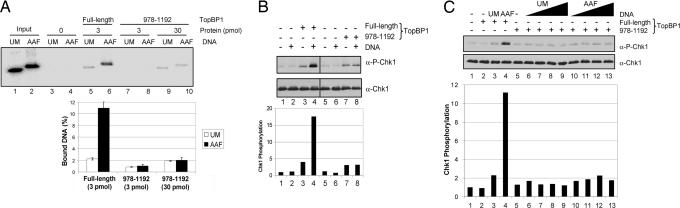
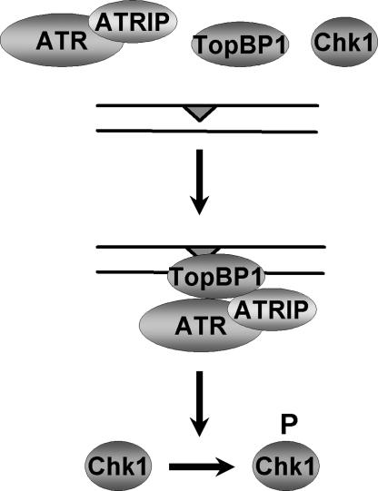
The DNA damage checkpoint response delays cell cycle progression upon DNA damage and prevents genomic instability. Genetic analysis has identified sensor, mediator, signal transducer, and effector components of this global signal transduction pathway. Here we describe an in vitro system with purified human checkpoint proteins that recapitulates key elements of the DNA damage checkpoint. We show that the damage sensor ATR in the presence of topoisomerase II binding protein 1 (TopBP1) mediator/adaptor protein phosphorylates the Chk1 signal-transducing kinase in a reaction that is strongly dependent on the presence of DNA containing bulky base lesions. The dependence on damaged DNA requires DNA binding by TopBP1, and, indeed, TopBP1 shows preferential binding to damaged DNA. This in vitro system provides a useful platform for mechanistic studies of the human DNA damage checkpoint response.


Conflict of interest statement

The authors declare no conflict of interest.

Figures

Fig. 1.
Fig. 1.
Purification of human ATR–ATRIP and activation of ATR kinase activity by TopBP1. (A) Scheme of ATR–ATRIP purification from HeLa cell-free extracts. (B) Purification of ATR–ATRIP. Fractions from the Mono S column were separated on a 5/10% discontinuous SDS/polyacrylamide gel and analyzed by Western blotting using the indicated antibodies (α-) (upper four blots) and by silver staining (lower gel). ATM and DNA-dependent PK catalytic subunit (DNAPKcs) were separated from fractions containing ATR–ATRIP during chromatography on CM Affi-Gel Blue and DEAE columns, respectively. The peak of ATR–ATRIP eluted at 220 mM KCl (fraction 8) from the Mono S column. NE, nuclear extract; L, load; E, eluate; FT, flow-through; 1–12, fraction numbers; asterisks, ATR and ATRIP. The line between the 100- and 150-kDa markers is the boundary between the 5% and 10% acrylamide regions of the gel. (C) Activation of purified ATR kinase activity by TopBP1. Kinase assays were performed with the Mono S column fractions to test for TopBP1-dependent ATR kinase activity. Mono S load (lanes 1 and 2), flow-through (lanes 3 and 4), and eluted fractions (lanes 5–12) were incubated under low ionic strength reaction conditions (45 mM total salt concentration) with His-Chk1-kd in the presence or the absence of GST-TopBP1-His. An equal volume of each fraction was preincubated for 15 min at 30°C with 5 nM GST-TopBP1-His. Chk1-kd (20 nM) was then added into the reaction and incubated for 20 min at 30°C. Reactions were analyzed by immunoblotting for phosphorylated Chk1 (P-Chk1, which is phosphorylated at S345) (Upper) and total Chk1 (Lower).
Fig. 2.
Fig. 2.
TopBP1-dependent stimulation of ATR kinase activity by DNA and effects of ionic strength on this stimulation. (Upper) Kinase assays were carried out with ATR–ATRIP, His-Chk1-kd, GST-TopBP1-His, and double-stranded DNA under different ionic strength reaction conditions. ATR–ATRIP (1 nM) was preincubated with or without 5 nM pUC19 plasmid in the presence and absence of 1 nM GST-TopBP1-His. Chk1-kd (20 nM) was then added to the reaction and incubated for 20 min at 30°C. (Lower) Levels of Chk1 phosphorylation were quantified, and the results from three independent experiments were averaged and graphed. Phosphorylation was normalized to the control reaction in lane 1 (without TopBP1 and DNA).
Fig. 3.
Fig. 3.
Stimulation of ATR kinase activity by various DNA substrates in the presence of TopBP1. (Upper) Kinase assays were performed with ATR–ATRIP, His-Chk1-kd, GST-TopBP1-His, and different forms of DNA substrates under high ionic strength reaction conditions (85 mM total salt concentration) as described in Materials and Methods. ATR–ATRIP (1 nM) was incubated with 1.25–5 nM single-stranded, deca-primed, or double-stranded pIBI25 plasmid in the presence and absence of 1 nM GST-TopBP1-His. (Lower) The average levels of Chk1 phosphorylation from three independent experiments were quantitated and graphed.
Fig. 4.
Fig. 4.
TopBP1-dependent stimulation of ATR kinase activity by N-Aco-AAF-damaged DNA. (A) (Upper) Kinase assays were performed with 85 mM final salt concentration as described in the legend of Fig. 3, except with 0.62–5 nM unmodified (UM) or N-Aco-AAF-damaged (AAF) pUC19 plasmid DNA. (Lower) The average levels of Chk1 phosphorylation from four independent experiments are quantitated in the graph. (B) (Upper) Time course of Chk1 phosphorylation. Kinase assays as in A were carried out with 0.62 nM DNA for the times indicated. (Lower) The average levels of Chk1 phosphorylation from two independent experiments are quantitated in the graph.
Fig. 5.
Fig. 5.
Dependence of DNA-binding activity of TopBP1 for ATR activation. (A) Preferential binding of TopBP1 to N-Aco-AAF-damaged DNA. (Upper) The indicated amounts of GST-TopBP1-His or the GST-TopBP1-(978–1192) fragment on glutathione-Sepharose beads was incubated at 37°C for 10 min with 1 ng of unmodified or N-Aco-AAF-damaged DNA that had been 5′-end-labeled. Bound DNA was eluted and analyzed by agarose gel electrophoresis. Fifty percent of the DNA added into the reaction was loaded in the input lanes. (Lower) Data from three independent experiments are presented as means ± SD in the graph. (B) Contribution of DNA-binding activity of TopBP1 to stimulation of ATR kinase activity by double-stranded DNA. (Upper) Kinase assays were performed as in Fig. 2, except with the TopBP1-(978–1192) fragment compared with full-length TopBP1 under low ionic strength reaction conditions (35 mM total salt concentration). The TopBP1-(978–1192) fragment (15 nM) was used to obtain a level of Chk1 phosphorylation comparable to that observed with 1 nM full-length TopBP1 (compare lanes 3 and 7). (Lower) The average levels of Chk1 phosphorylation from two independent experiments are quantitated in the graph. (C) Requirement of DNA-binding activity of TopBP1 for stimulation of ATR kinase activity by N-Aco-AAF-damaged DNA. (Upper) Kinase assays were performed with 85 mM final salt concentration as in Fig. 4A, except with the TopBP1-(978–1192) fragment (15 nM) compared with full-length TopBP1 (1 nM). For the reactions containing full-length TopBP1, 5 nM unmodified or N-Aco-AAF-damaged DNA was added. (Lower) The average levels of Chk1 phosphorylation from two independent experiments are quantitated in the graph.
Fig. 6.
Fig. 6.
Model of TopBP1-dependent stimulation of ATR kinase activity by damaged DNA. Data from this study suggest that bulky DNA lesions are recognized by TopBP1, which recruits ATR to the damage site and potentiates its kinase activity on the checkpoint signal transduction kinase, Chk1.

Similar articles

Cited by

References

    1. Abraham RT. Genes Dev. 2001;15:2177–2196. - PubMed
    1. Nyberg KA, Michelson RJ, Putnam CW, Weinert TA. Annu Rev Genet. 2002;36:617–656. - PubMed
    1. Sancar A, Lindsey-Boltz LA, Ünsal-Kaçmaz K, Linn S. Annu Rev Biochem. 2004;73:39–85. - PubMed
    1. Lee JH, Paull TT. Science. 2004;304:93–96. - PubMed
    1. Lee JH, Paull TT. Science. 2005;308:551–554. - PubMed

Publication types