. 2007 Oct;18(10):3952-65.
doi: 10.1091/mbc.e07-07-0678. Epub 2007 Aug 8.

Plasticity of polyubiquitin recognition as lysosomal targeting signals by the endosomal sorting machinery

Affiliations

Plasticity of polyubiquitin recognition as lysosomal targeting signals by the endosomal sorting machinery

Herve Barriere et al. Mol Biol Cell. 2007 Oct.

Abstract

Lysosomal targeting is fundamental for the regulated disposal of ubiquitinated membrane proteins from the cell surface. To elucidate ubiquitin (Ub) configurations that are necessary and sufficient as multivesicular body (MVB)/lysosomal-sorting motifs, the intraendosomal destination and transport kinetics of model transmembrane cargo molecules bearing monoubiquitinated, multi-monoubiquitinated, or polyubiquitinated cytoplasmic tails were determined. Monomeric CD4 chimeras with K63-linked poly-Ub chains and tetrameric CD4-mono-Ub chimeras were rapidly targeted to the lysosome. In contrast, lysosomal delivery of CD4 chimeras exposing K48-linked Ub chains was delayed, whereas delivery of monoubiquitinated CD4 chimeras was undetectable. Similar difference was observed in the lysosomal targeting of mono- versus polyubiquitinated invariant chain and CD4 ubiquitinated by the MARCH (membrane-associated RING-CH) IV Ub ligase. Consistent with this, Hrs (hepatocyte growth factor regulated tyrosine kinase phosphorylated substrate), an endosomal sorting adaptor, binds preferentially to K63-Ub chain and negligibly to mono-Ub. These results highlight the plasticity of Ub as a sorting signal and its recognition by the endosomal sorting machinery, and together with previous data, suggest a regulatory role for assembly and disassembly of Ub chains of specific topology in lysosomal cargo sorting.


Figures
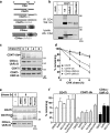
Figure 1.
Figure 1.
Expression and stability of CD4-Ub chimeras. (a) Schematic representations CD4-Ub chimeras. The cytoplasmic tail of CD4 was replaced with a flexible linker (CD4Tl) or a tetramerization coiled-coil domain (CD4cc) and the indicated Ub moiety as described in Materials and Methods. (b) Polyubiquitination of the CD4Tl-Ub. COS-7 transiently cell expressing the indicated CD4 chimeras, were incubated with OKT4 anti-CD4 antibody for 30 min at 37°C. CD4-Ab complexes were immunoisolated on protein G beads. Immunoprecipitates were probed with HRP-conjugated P4D1 anti-Ub antibody. (c) Metabolic stability of the CD4 chimeras was measured after inhibiting translation with 100 μg/ml cycloheximide (CHX) in transiently transfected HEK293 cells. After the chase, equal amounts of cell lysates were immunoblotted with anti-CD4 Ab. Ubiquitination of CD4Tl-Ub is indicated by an asterisk (*). (d) Degradation kinetics of CD4-Ub chimera were determined by densitometry of immunoblots illustrated in panel c. Data are means ± SEM; n = 3–4. (e) Inhibition of lysosomal proteolysis and cargo delivery stabilizes CD4Tl-Ub and CD4cc-UbRΔG. The stability of CD4-Ub chimeras was measured in the presence of bafilomycinA1 (1 μM), MG132 (20 μM), or leupeptin+pepstatin (5–5 μg/ml) as described in panel c after a 6-h CHX chase. (f) Densitometric analysis of CD4Tl, CD4Tl-Ub, and CD4cc-UbRΔG immunoblots as shown in panel e. Lactacystin at 10 μM was used. Data are expressed as percentage of the initial amount of cargo. Means ± SEM; n = 3.
Figure 2.
Figure 2.
Stability, internalization and recycling of CD4-Ub chimeras. (a) The turnover of plasma membrane pool of chimeras was monitored by the disappearance of cell surface bound anti-CD4 Ab in transiently transfected COS-7 cells. Cells were labeled with anti-CD4 Ab on ice for 1 h and then chased for 0.5–20 h at 37°C. The Ab remaining at the cell surface was measured by fluorescence in the presence of HRP-conjugated secondary Ab and AmplexRed, as described in Materials and Methods. The fluorescence was normalized for cellular proteins. Means ± SEM; n = 3. (b) Internalization rates of CD4-Ub chimeras. Endocytic rates of chimeras were measured by anti-CD4 Ab uptake in COS-7 cells as described in Materials and Methods. Data are expressed as percentage of the initial amount of CD4-Ub chimeras at the cell surface. Means ± SEM; n = 3–5. (c) Recycling efficiency of chimeras was measured in transiently transfected COS-7 cells with the biotin-streptavidin sandwich technique as described in Materials and Methods. Recycling was expressed as the percentage of internalized cargo. Means ± SEM; n = 3–5.
Figure 3.
Figure 3.
Subcellular localization of internalized CD4-Ub chimeras. (a) Internalized CD4Tl-Ub and CD4cc-UbRΔG colocalizes with lysosomes and avoid recycling endosomes in HEK293 cells. Chimeras were internalized with anti-CD4 Ab and FITC-conjugated secondary Fab. Lysosomes and recycling endosomes were labeled with FITC-dextran and TRITC-Tf, respectively. Internalized dextran was colocalized with Lamp1 by immunostaining (data not shown). Single optical sections were obtained by laser confocal fluorescence microscopy. Bar, 10 μm. (b) Internalized CD4Tl and CD4Tl-UbRΔG colocalize with recycling endosomes and excluded from lysosomes. Staining of recycling endosomes, lysosomes, and CD4 chimeras were performed as described in panel a. Bar, 10 μm.
Figure 4.
Figure 4.
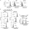
Monitoring internalized cargo sorting by vesicular pH (pHv) measurement. (a) Validation of pHv measurements by monitoring transferrin (Tf), EGF (epidermal growth factor), and dextran sorting in HEK293 cells. The loading protocols of FITC-dextran and FITC–Tf are described in Materials and Methods. Serum-depleted cells internalized biotin-EGF and FITC-streptavidin for 1 h and chased for 2 h at 37°C. The pHv was measured by FRIA. Similar results were obtained in COS-7 (dextran) cells. The mean (±SEM) pHv and the number of vesicles analyzed in a single experiment are indicated. The mean pHv from ≥3 independent experiments are summarized in Supplementary Table S1. (b) The kinetic of CD4Tl-Ub lysosomal delivery. Anti-CD4 antibody and FITC-conjugated secondary Fab was bound to transfected HEK293 cells for 1 h at 0°C. Then the temperature was raised to 37°C for 1, 15, or 30 min, and the pHv was measured by FRIA. (c) CD4Tl- and CD4Tl-Ub–expressing HEK293 cells were allowed internalize anti-CD4 primary Ab and FITC-conjugated secondary Fab for 1 h at 37°C and were chased for 30 min before FRIA. Similar results were obtained in COS-7 cells (data not shown).
Figure 5.
Figure 5.
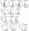
Lysosomal sorting of CD4-Ub chimera requires poly-Ub or multimeric-Ub. (a) Postendocytotic sorting of the indicated CD4 chimera in COS-7 cells was monitored by FRIA as described in Figure 4c. Data are expressed as frequency of pHv and means of pHv ± SEM from a single experiment (see also Supplementary Table S1). (b) Lysosomal sorting of invariant chain-Ub chimeras. The postendocytic trafficking of the truncated invariant chain (IiT) lacking its internalization motif, IiT-Ub and IiT-UbRΔG was determined by the pHv measurement using primary anti-Ii– and FITC–conjugated secondary Fab fragment with FRIA as described in Materials and Methods. (c) The pHv of internalized CD4cc-, CD4cc-Ub–, and CD4cc-UbRΔG–containing vesicles was measured as in Figure 4c. (d). Multimerization of CD4Tl and CD4Tl-UbRΔG was achieved by incubating the cells sequentially with anti-CD4 Ab (OKT4), biotinylated secondary Ab and streptavidin-FITC on ice. Internalization was initiated at 37°C in Ab-free medium for 30 min, and pHv was measured by FRIA. (e) Postendocytic sorting of CD4cc-UbRI44A and CD4cc-UbR was established by pHv measurements as in panel a.
Figure 6.
Figure 6.
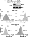
Inactivation of the E1 enzyme impairs Ub chain formation and lysosomal sorting of the CD4Tl-Ub, but not the CD4cc-UbRΔG. (a) Thermoinactivation of the E1 enzyme was accomplished at 40°C in ts20, but not in E36 cells. Equal amounts of cell lysates were probed with anti-E1 Ab using ECL. (b) Ts20 and E36 cells, expressing the indicated construct, were incubated at 40°C for 3 h to down-regulate the E1 Ub-activating enzyme. The pHv of internalized CD4Tl-Ub and CD4cc-UbRΔG was determined by FRIA.
Figure 7.
Figure 7.
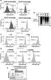
The effect of Ub chain configuration on the internalization and postendocytic sorting of CD4-Ub chimeras. (a) Postendocytic sorting of CD4Tl-UbRΔG6K, -UbRΔG29K, -UbRΔG48K, and -UbRΔG63K was monitored by FRIA in transiently transfected COS-7 cells as in Figure 4c. The vesicles displaying the larger cohort of pHv have gray shade. (b) Immunoblot analysis of COS-7 cells transiently transfected with HA-tagged Ub variants. Equal amounts of cell lysates were separated by SDS-PAGE and probed with anti-HA Ab. (c) Transient coexpression of UbR, but not wt Ub, prevents lysosomal accumulation of CD4Tl-Ub. The postendocytic fate of chimeras was monitored by pHv as in Figure 4c in COS-7 cells transiently coexpressing CD4TlUb and Ubs variants at a plasmid ratio of 1:3–4. (d) The postendocytic sorting of CD4Tl-Ub in the presence of overexpressed UbR63K or UbR48K was monitored by pHv. When indicated, the chase was extended to 90 min. Similar results were obtained in HEK293 cells stably expressing CD4Tl-Ub and transfected with plasmids encoding the Ub variant and the dsRed fluorescent protein at a ratio of 10:1. FRIA was performed on cell with dsRed expression. (e) Internalization rates of CD4Tl-Ub in the presence of overexpressed Ub variant. Endocytic rates were measured by the anti-CD4 Ab uptake assay in transiently cotransfected COS-7 cells as described in Materials and Methods. Means ± SEM; n = 3; *p > 0.05 relative to mock transfected CD4Tl-Ub.
Figure 8.
Figure 8.
Ub binding specificity of endogenous and recombinant Hrs. (a) Association of Hrs with CD4Tl-Ub in HEK293 cells. Immunoprecipitated CD4Tl-Ub with the OKT4 anti-CD4 Ab was probed with the indicated Ab. One hundred micrograms of lysates (lys) was loaded. (b) Equal amounts of GST fusion proteins containing one, two, three, or four tandem Ubs were incubated with HeLa cell lysate as described in Materials and Methods. Bound Hrs (top panel) and E1 Ub-activating enzyme (middle panel) was visualized by immunoblotting. The mutant variants of GST-2Ub are described in Materials and Methods. Ten percent of lysate (lys) was loaded. GST-Ub fusions are visualized by Ponceau staining (bottom panel). (c) Preferential binding of K63-linked Ub-chain binding to recombinant Hrs. Similar amounts of MBP, wt, or mutant MBP-Hrs fusion proteins were bound to amylose beads and incubated with K48- (left panel), K63-linked poly-Ub chain (middle panel), or mono-Ub (right panel). Bound Ub was visualized by immunoblotting, using the P4D1 anti-Ub Ab. Ubs were also loaded for direct comparison as specified in Materials and Methods.
Figure 9.
Figure 9.
Polyubiquitination is required for CD4-1K lysosomal targeting. (a) Postendocytic sorting of CD4-1K and CD4-4R in the presence or absence of MARCH-IV and wt Ub was monitored by FRIA in transiently transfected COS-7 cells. Abs were internalized for 1.5 h and chased for the indicated time. (b) Postendocytic sorting of CD4-1K was monitored in the presence of MARCH-IV and UbR, UbR63K, or UbR48K as described in panel a. The chase period was 45 or 90 min.

References

    1. Amerik A. Y., Hochstrasser M. Mechanism and function of deubiquitinating enzymes. Biochim. Biophys. Acta. 2004;1695:189–207. - PubMed
    1. Aniento F., Gu F., Parton R. G., Gruenberg J. An endosomal beta COP is involved in the pH-dependent formation of transport vesicles destined for late endosomes. J. Cell Biol. 1996;133:29–41. - PMC - PubMed
    1. Bache K. G., Raiborg C., Mehlum A., Stenmark H. STAM and Hrs are subunits of a multivalent ubiquitin-binding complex on early endosomes. J. Biol. Chem. 2003;278:12513–12521. - PubMed
    1. Barriere H., Nemes C., Lechardeur D., Khan-Mohammad M., Fruh K., Lukacs G. L. Molecular basis of oligoubiquitin-dependent internalization of membrane proteins in mammalian cells. Traffic. 2006;7:282–297. - PubMed
    1. Bartee E., Mansouri M., Hovey Nerenberg B. T., Gouveia K., Fruh K. Downregulation of major histocompatibility complex class I by human ubiquitin ligases related to viral immune evasion proteins. J. Virol. 2004;78:1109–1120. - PMC - PubMed

Publication types

MeSH terms