Comparative Study
. 2007 Aug 8;27(32):8654-64.
doi: 10.1523/JNEUROSCI.1615-07.2007.

Functional properties of neurons derived from in vitro reprogrammed postnatal astroglia

Affiliations
Comparative Study

Functional properties of neurons derived from in vitro reprogrammed postnatal astroglia

Benedikt Berninger et al. J Neurosci. .

Abstract

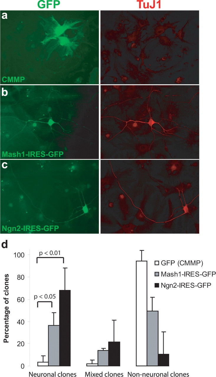
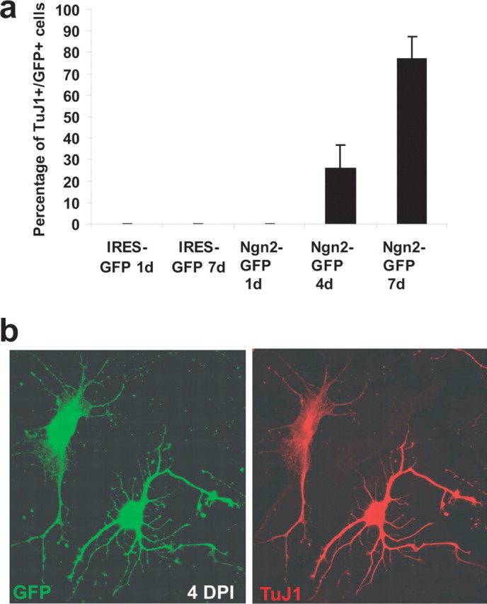
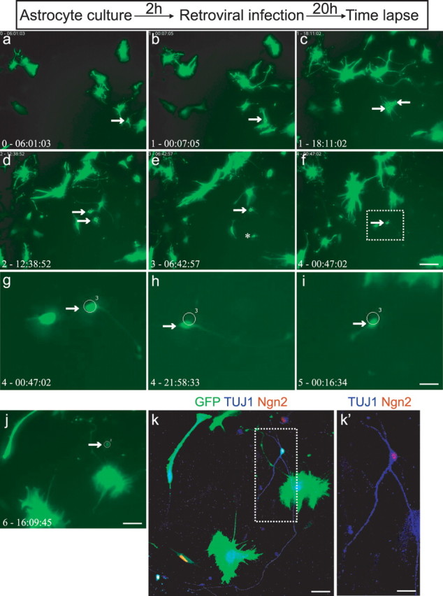
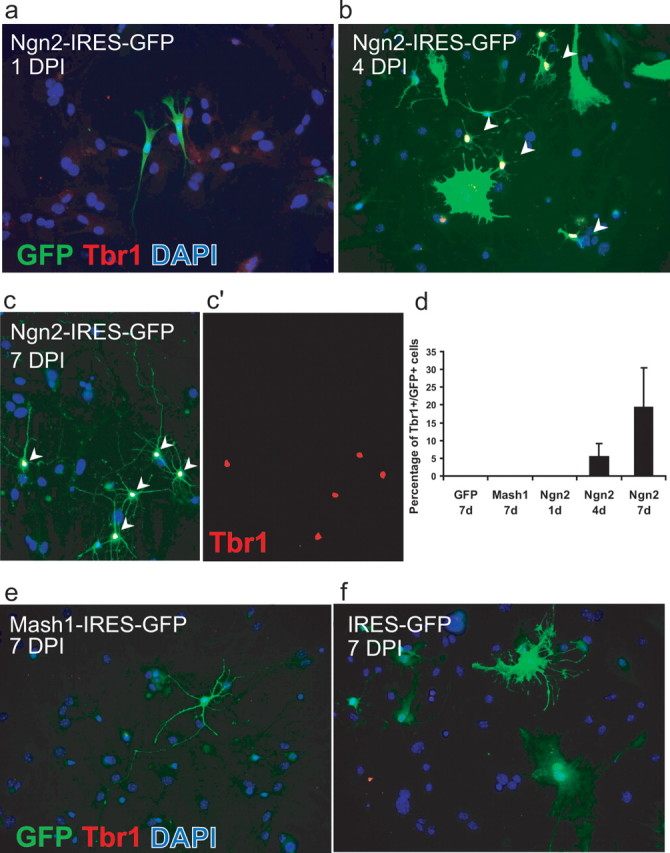
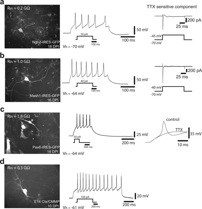
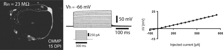
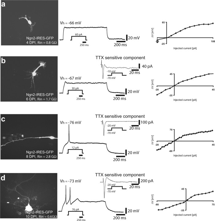
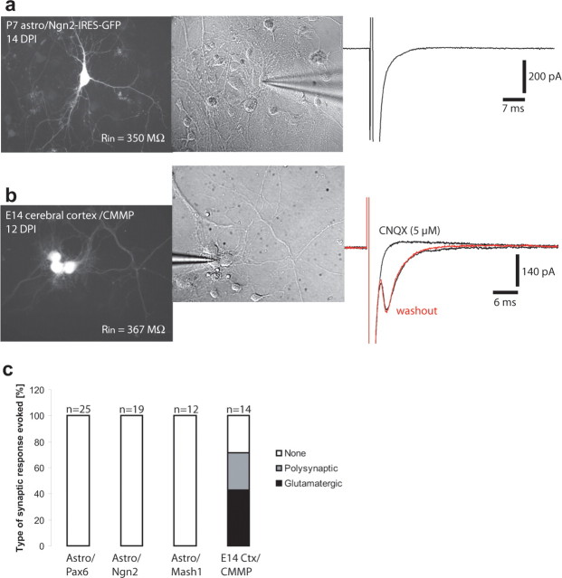
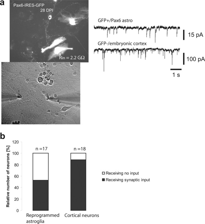
With the exception of astroglia-like cells in the neurogenic niches of the telencephalic subependymal or hippocampal subgranular zone, astroglia in all other regions of the adult mouse brain do not normally generate neurons. Previous studies have shown, however, that early postnatal cortical astroglia in culture can be reprogrammed to adopt a neuronal fate after forced expression of Pax6, a transcription factor (TF) required for proper neuronal specification during embryonic corticogenesis. Here we show that also the proneural genes neurogenin-2 and Mash1 (mammalian achaete schute homolog 1) possess the ability to reprogram astroglial cells from early postnatal cerebral cortex. By means of time-lapse imaging of green fluorescent astroglia, we provide direct evidence that it is indeed cells with astroglial characteristics that give rise to neurons. Using patch-clamp recordings in culture, we show that astroglia-derived neurons acquire active conductances and are capable of firing action potentials, thus displaying hallmarks of true neurons. However, independent of the TF used for reprogramming, astroglia-derived neurons appear to mature more slowly compared with embryonic-born neurons and fail to generate a functional presynaptic output within the culturing period. However, when cocultured with embryonic cortical neurons, astroglia-derived neurons receive synaptic input, demonstrating that they are competent of establishing a functional postsynaptic compartment. Our data demonstrate that single TFs are capable of inducing a remarkable functional reprogramming of astroglia toward a truly neuronal identity.


Figures

Figure 1.
Figure 1.
Ngn2 and Mash1 can reprogram astroglial cells to adopt a neuronal fate. a, Examples of astroglia transduced with a vector only encoding GFP (CMMP) 12 d after infection. Left, GFP immunoreactivity; right, TuJ1 immunoreactivity. b, Example of Mash1-transduced cell also positive for TUJ1 12 d after infection. c, Example of Ngn2-transduced cell double-positive for TUJ1 12 d after infection. d, Summary of three independent experiments in which the number of clones consisting of TuJ1-positive (neuronal), TuJ1-positive as well as -negative (mixed), and only TuJ1-negative (non-neuronal) cells is given as a percentage of all clones analyzed. Cells were fixed 12 d after infection and analyzed in three different experimental batches with >100 cells counted in each batch. For statistical analysis, a nonparametric ANOVA was used with a Dunn's post hoc analysis.
Figure 2.
Figure 2.
Gradual acquisition of neuronal markers by cells of astroglial origin during reprogramming with Ngn2. a, Time course of TuJ1 immunoreactivity in astroglial cultures transduced with Ngn2. b, Example of two cells expressing Ngn2–IRES–GFP (green, left) 4 d after transduction. Both cells express TuJ1 (red, right). Note the partially astroglial morphology of the left cell indicating a transitional state.
Figure 3.
Figure 3.
Time-lapse video microscopy of hGFAP–GFP-positive cells transforming into neurons after Ngn2 transduction. a–j, Time-lapse images of an astroglia culture derived from a P7 hGFAP–GFP transgenic mouse after transduction with Ngn2. Arrows point to an GFP-positive astroglial cell at an early stage when virally mediated GFP expression is not yet detectable (a, b). The progeny of this cell is delineated by two arrows (c, d) shortly after cell division. The daughter cell depicted by an asterisk (e) then undergoes cell death, whereas the remaining cell (depicted at higher power in g–j) underwent a marked change in cell morphology, displaying neuronal-like characteristics (arrows in f–j). The corresponding time is indicated in the left corner of each image (in days-hours:minutes:seconds). Box in f is amplified in g–i. k, k′, After imaging, cells were fixed and immunocytochemically analyzed for GFP (green), Tuj1 (blue), and Ngn2 (red). The neuronal identity of the reprogrammed cell was confirmed by its immunoreactivity for TuJ1. Box in k is shown at higher magnification in k′. Note the Ngn2 immunoreactivity in the nucleus, confirming that the neuron was derived from a Ngn2-transduced cell. Scale bars: a–f, j, 120 μm; k, 50 μm; g–i, 30 μm; k′, 25 μm.
Figure 4.
Figure 4.
Upregulation of the T-box transcription factor Tbr1 after reprogramming of astroglial cells by Ngn2 but not Mash1. a–c, Representative images taken at discrete stages of maturation after transduction with Ngn2. a, Depicted are two GFP-positive (green)/Tbr1-negative (red) cells 1 d after transduction with Ngn2–IRES–GFP. b, By day 4, some Ngn2-expressing cells with a neuroblast morphology exhibit Tbr1 immunoreactivity (white nuclei depicted by arrowheads). c, c′, By day 7, an additional increased number of Ngn2-transduced cells is also Tbr1 positive (white nuclei indicated by arrowheads in c and red nuclei in c′). Most of these cells now display a distinctly neuronal morphology. Cells were stained for GFP (green) to visualize transduced cells, Tbr1 (red), and 4′,6′-diamidino-2-phenylindole (DAPI) to identify nuclei. d, Quantification of Tbr1-positive cells after transduction with control vector, Mash1, or Ngn2. Only the 7 d time point is shown for control or Mash1 transduction, but also at previous stages no Tbr1 immunoreactivity was found. In the case of Ngn2, a gradual increase in the number of Tbr1-positive cells was noted. e, Seven days after transduction with Mash1, neurons did not exhibit Tbr1 immunoreactivity. f, Control transduced cells were Tbr1 negative.
Figure 5.
Figure 5.
Astroglial cells reprogrammed by neurogenic fate determinants fire action potentials. a, Example of a neuron derived from an astroglial cell reprogrammed by Ngn2 18 DPI. Left shows cell expressing Ngn2–IRES–GFP. Step current injection (50 pA for 300 ms) in current clamp resulted in repetitive firing of action potentials (middle). The right shows the TTX-sensitive Na+ current induced by step depolarization from −70 to −45 mV. The trace shown was obtained by subtracting the current response in the presence of 1 μm TTX from the control trace. b, Example of a neuron derived from an astroglial cell reprogrammed by Mash1 (16 DPI). Left shows expression of GFP after transduction with Mash1–IRES–GFP. The middle shows the repetitive discharge of action potentials in response to current injection in current clamp. The right shows the TTX-sensitive Na+ current activated by step depolarization from −70 to −40 mV. c, Example of a neuron (marked by asterisk) derived from an astroglial cell reprogrammed by Pax6. The left shows expression of GFP after transduction with Pax6–IRES–GFP. The middle shows the repetitive spiking behavior of the cell after current injection. The right shows that action potentials were blocked by 1 μm TTX. d, Example of a neuron derived from embryonic cortex and transduced with a control vector encoding GFP only (CMMP). The cell exhibited a repetitive firing in response to current injection in current clamp. Vh indicates the membrane potential at which the cells had been kept.
Figure 6.
Figure 6.
Control GFP virus-transduced astroglia do not fire action potentials. Example of a cell transduced with virus containing only GFP (see micrograph; for construct, see Materials and Methods), exhibiting a linear current–voltage relationship. Middle shows a family of membrane potentials in response to step current injection. Right shows voltage changes in response to current injection.
Figure 7.
Figure 7.
Gradual acquisition and maturation of action potential firing in neurons derived from Ngn2 reprogrammed astroglia. This series of recordings were performed on the same culture preparation to allow comparable insight into neuronal maturation of reprogrammed astrocytes. a, Example of a cell expressing Ngn2–IRES–GFP for a period of 4 d. Depolarizing current injection did not yet evoke a spike or spikelet (middle). However, the current–voltage relationship (right) revealed an increase in input resistance during large current injections indicative of active conductances. b, Example of a cell expressing Ngn2–IRES–GFP for a period of 6 d. Depolarizing current injection evoked a small spikelet in current clamp. In voltage-clamp activation of small Na+ current component could be revealed by subtracting the current response in TTX from the control trace (middle). The current–voltage relationship was still fairly passive. c, Example of a cell expressing Ngn2–IRES–GFP for a period of 8 d (left). The cell fired a single action potential after current injection in current clamp (middle). In voltage clamp, a markedly increased Na+ current component could be revealed by block with TTX. The overall current–voltage relationship exhibited a slightly more nonlinear behavior compared with the cell shown in b. d, Example of a cell expressing Ngn2–IRES–GFP for a period of 10 d. The cell fired a doublet of spikes in response to current injection, which correlated with a peak Na+ current of ∼600 pA. The current–voltage relationship exhibited nonlinearity in both the hyperpolarizing and depolarizing range. Vh, Holding potential.
Figure 8.
Figure 8.
Neurons derived from reprogrammed postnatal astroglial precursors do not establish functional autapses. a, Example of an Ngn2-transduced cell. Left, A GFP-positive neuron. Middle, The same cell under bright field, and the right shows absence of autaptic currents after step depolarization of the neuron. b, Example of a clone of neurons obtained from E14 embryonic cerebral cortex and transduced with a GFP only encoding retrovirus (CMMP). The recorded neuron exhibited a glutamatergic autapse as indicated by the total block of the response by CNQX. After washout of CNQX the response recovered. c, Summary of the type of responses that could be evoked from cells derived from P5–P7 astroglial precursors reprogrammed by Pax6, Ngn2, or Mash1 10–28 d after infection and CMMP-transduced neurons from E14 cerebral cortex cultures kept for 10–21 d in vitro. Evoked responses were observed only in the latter.
Figure 9.
Figure 9.
Reprogrammed astroglial cells receive functional synaptic input. a, Example of a dual recording of a neuron derived from a Pax6 reprogrammed cell and a cocultured neuron from E16 cerebral cortex. Both neurons receive synaptic input, but the frequency and amplitude of synaptic events in the glia-derived cell are notably lower compared with the E16 cortical neuron. b, Percentage of neurons derived from reprogrammed astroglial precursors and cocultured cortical neurons receiving synaptic input in coculture.

Similar articles

Cited by

References

    1. Ackman JB, Siddiqi F, Walikonis RS, LoTurco JJ. Fusion of microglia with pyramidal neurons after retroviral infection. J Neurosci. 2006;26:11413–11422. - PMC - PubMed
    1. Anderson S, Mione M, Yun K, Rubenstein JLR. Differential origins of neocortical projection and local circuit neurons: role of Dlx genes in neocortical interneuronogenesis. Cereb Cortex. 1999;9:646–654. - PubMed
    1. Bachoo RM, Maher EA, Ligon KL, Sharpless NE, Chan SS, You MJ, Tang Y, DeFrances J, Stover E, Weissleder R, Rowitch DH, Louis DN, DePinho RA. Convergent mechanisms governing terminal differentiation and transformation along the neural stem cell to astrocyte axis. Cancer Cell. 2002;1:269–277. - PubMed
    1. Ballas N, Grunseich C, Lu DD, Speh JC, Mandel G. REST and its corepressors mediate plasticity of neuronal gene chromatin throughout neurogenesis. Cell. 2005;121:645–657. - PubMed
    1. Berninger B, Guillemot F, Götz M. Directing neurotransmitter identity of neurons derived from expanded adult neural stem cells. Eur J Neurosci. 2007;25:2581–2590. - PubMed

Publication types