Comparative Study
. 2007 Aug 8;27(32):8676-86.
doi: 10.1523/JNEUROSCI.0658-07.2007.

Munc18-1: sequential interactions with the fusion machinery stimulate vesicle docking and priming

Affiliations
Comparative Study

Munc18-1: sequential interactions with the fusion machinery stimulate vesicle docking and priming

Attila Gulyás-Kovács et al. J Neurosci. .

Abstract

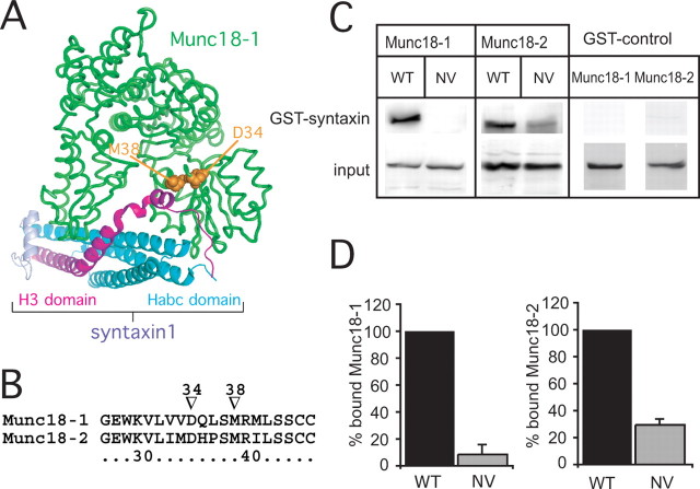
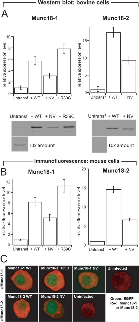
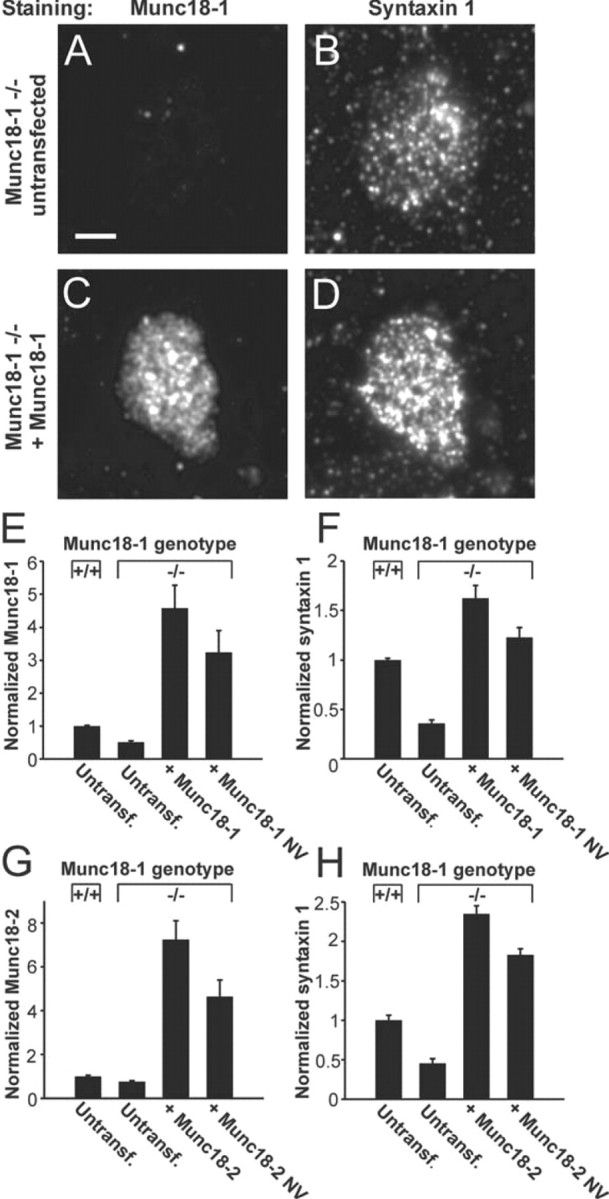
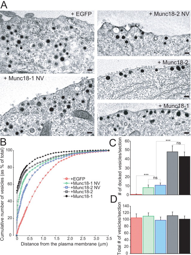
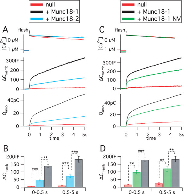
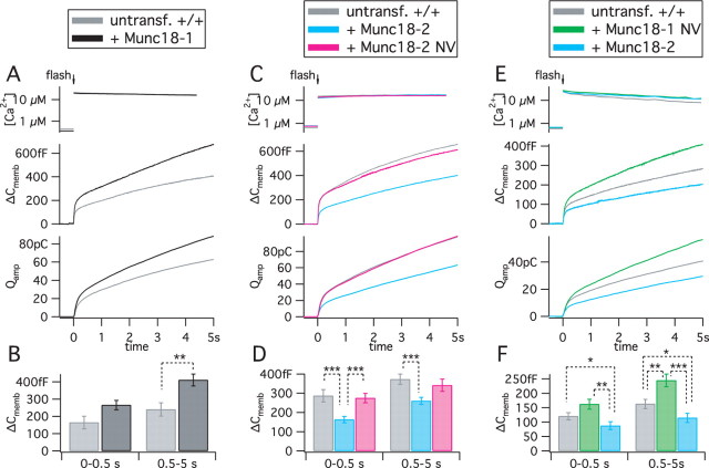
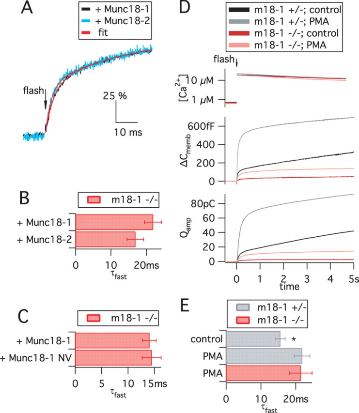
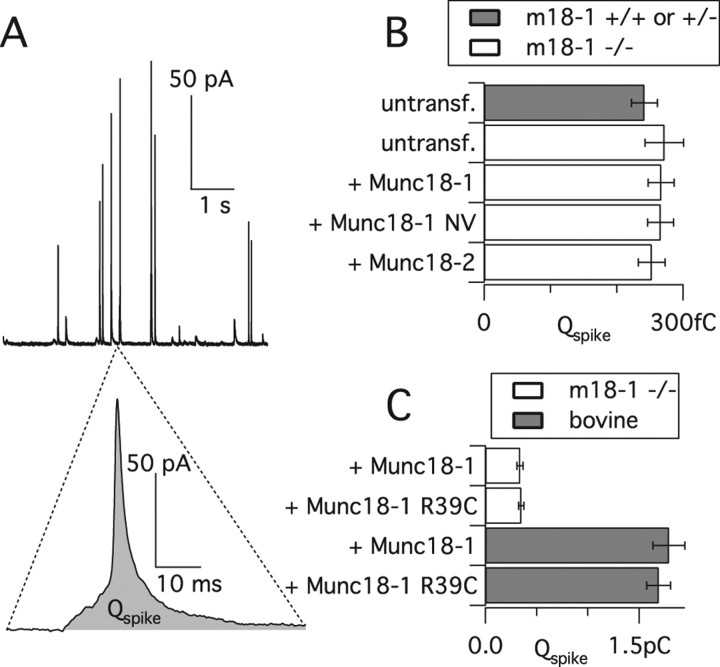
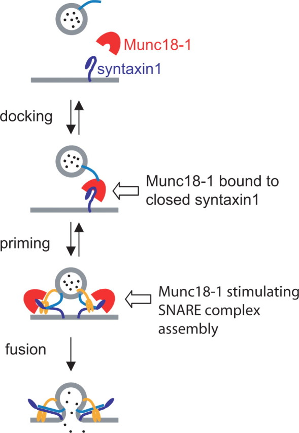
Exocytosis of secretory or synaptic vesicles is executed by a mechanism including the SNARE (soluble N-ethylmaleimide-sensitive factor attachment protein receptor) proteins. Munc18-1 is a part of this fusion machinery, but its role is controversial because it is indispensable for fusion but also inhibits the assembly of purified SNAREs in vitro. This inhibition reflects the binding of Munc18-1 to a closed conformation of the target-SNARE syntaxin1. The controversy would be solved if binding to closed syntaxin1 were shown to be stimulatory for vesicle fusion and/or additional essential interactions were identified between Munc18-1 and the fusion machinery. Here, we provide evidence for both notions by dissecting sequential steps of the exocytotic cascade while expressing Munc18 variants in the Munc18-1 null background. In Munc18-1 null chromaffin cells, vesicle docking is abolished and syntaxin levels are reduced. A mutation that diminished Munc18 binding to syntaxin1 in vitro attenuated the vesicle-docking step but rescued vesicle priming in excess of docking. Conversely, expressing the Munc18-2 isoform, which also displays binding to closed syntaxin1, rescued vesicle docking identical with Munc18-1 but impaired more downstream vesicle priming steps. All Munc18 variants restored syntaxin1 levels at least to wild-type levels, showing that the docking phenotype is not caused by syntaxin1 reduction. None of the Munc18 variants affected vesicle fusion kinetics or fusion pore duration. In conclusion, binding of Munc18-1 to closed syntaxin1 stimulates vesicle docking and a distinct interaction mode regulates the consecutive priming step.


Figures

Figure 1.
Figure 1.
The NV mutation in Munc18-1 and Munc18-2 diminishes syntaxin1 binding. A, Structural model of Munc18-1 in complex with the closed conformation of syntaxin1 (Misura et al., 2000) (Protein Data Bank code 1DN1). The mutated residues of Munc18-1 NV are shown in orange filled representation. B, Sequence alignment of a short fragment of Munc18-1 and Munc18-2. Arrowheads point to the mutated residues in the NV mutants. C, GST–syntaxin1 binding of Munc18-1 and Munc18-2 (WT) and their NV mutants. D, Mean ± SEM binding of Munc18-1 NV and Munc18-2 NV to syntaxin1 as a fraction of WT binding. n = 4 experiments.
Figure 2.
Figure 2.
Protein levels of native and overexpressed Munc18 s in adrenal chromaffin cells. A, Quantification (top row) and representative Western blots (bottom row) stained for Munc18-1 (left column) or Munc18-2 (right column) from bovine cells (3 μg protein/lane). Cells were infected with SFV expressing the WT, NV, or R39C variant of Munc18s. For uninfected cells, 10 times the amount (30 μg protein/lane) was loaded to illustrate native Munc18 expression. The quantification of Munc18-1 and Munc18-2 protein levels was corrected for infected efficiency (40–80% of the cells were expressing the different constructs). n = 3 cell preparations. B, Quantification of protein levels from expressing and untransfected embryonic mouse chromaffin cells (munc18-1 +/+) using immunofluorescence. The NV mutations were expressed at half the level of wild-type proteins. Data are mean ± SEM from 29–51 cells. C, Confocal sections through the equatorial plane of embryonic mouse chromaffin cells (munc18-1 +/+) immunostained for Munc18-1 (top row) or Munc18-2 (bottom row) and expressing the constructs indicated. Red, Munc18-specific staining; green, EGFP fluorescence. EGFP was expressed from the same viral constructs as a separate protein and found throughout the cell but is visible here mainly in the nucleus because of the intense cytoplasmic Munc18 staining. Munc18-1 and Munc18-2 were found throughout the cytoplasm, as expected. In addition, in some but not all cells, Munc18-2 NV was found concentrated in spots within the cytosol. Refer also to Figure 3, which shows that all variants were present on plasma membrane sheets. Note that the confocal sections were taken with different photomultiplier settings to visualize the distribution and therefore do not yield quantitative information about expression levels. Quantitative information is present in A and B.
Figure 3.
Figure 3.
Munc18-1 and its variants stabilize syntaxin1 on the plasma membrane. A–D, Immunodetection of Munc18-1 (A, C) and syntaxin1 (B, D) in isolated plasma membrane sheets of munc18-1 −/− cells. After expression of Munc18-1, sheets from −/− cells show increased staining with the syntaxin1 antibody (D vs B) and with the Munc18-1 antibody (C vs A). Scale bar, 3 μm. E, F, Quantification of Munc18-1 (E) and syntaxin1 (F) immunofluorescence from membrane sheets of +/+ cells or −/− cells expressing Munc18-1 or Munc18-1 NV. Note that, even in untransfected −/− cells, a fluorescence signal in the Munc18-1-specific channel was detected, indicating some unspecific binding of the Munc18-1 antibody. G, H, Immunofluorescence for Munc18-2 (G) and syntaxin1 (H) at the plasma membrane after overexpression of Munc18-2 or Munc18-2 NV. Note that NV mutants of both Munc18 isoforms were able to rescue syntaxin1 level in −/− cells to above the level in untransfected +/+ cells.
Figure 4.
Figure 4.
Stimulatory role of Munc18-1 binding to closed syntaxin1 in the docking step. A, The electron micrographs show the intracellular distribution of large dense-core vesicles close to the plasma membrane. munc18-1 −/− (null) cells expressed EGFP with or without variants of Munc18 proteins. Note that vesicles are not in close contact with the plasma membrane without Munc18s. Scale bars, 100 nm. B, Normalized cumulative distribution of vesicles as a function of distance from the plasma membrane. Data represent several cells/condition (see below). C, D, The number of docked vesicles (C) and the total number of vesicles (D). The data show that the ability of Munc18 to bind to syntaxin1 correlates with the level of rescue of vesicle docking. Data are mean ± SEM from the following number of cells (n) and animals (N): EGFP, n = 19, N = 4; Munc18-1 NV, n = 22, N = 7; Munc18-2 NV, n = 20, N = 3; Munc18-2, n = 20, N = 3; Munc18-1, n = 18, N = 3. ANOVA followed by Tukey–Kramer post hoc test. All conditions are significantly different at ***p < 0.001, except Munc18-1NV versus Munc18-2 NV and Munc18-1 versus Munc18-2, which are statistically identical.
Figure 5.
Figure 5.
Differential priming by isoforms reveals a second interaction between Munc18-1 and the fusion machinery. A, Release of primed vesicles after rapid Ca2+ uncaging in munc18-1 −/− cells expressing Munc18-1 or Munc18-2. Untransfected −/− (null) cells were used as control. The top graph shows the increases of intracellular [Ca2+] by the flash of UV light. This evokes rapid membrane fusion and concomitant catecholamine release from primed vesicles in the burst phase, followed by replenishment of the primed pool in the sustained phase. Fusion is assayed by membrane capacitance measurements (ΔC memb) and catecholamines are detected by amperometry (Q amp). Traces are averages from n cells: n = 12 untransfected; n = 31 Munc18-1 and 30 Munc18-2. B, Mean ± SEM representation of the burst (0–0.5 s) and sustained (0.5–5 s) phases from the membrane capacitance responses. ***p < 0.0001. C, D, Munc18-1 NV was evaluated similarly to Munc18-2 above. n = 5 untransfected cells; n = 36 Munc18-1 NV expressing cells; and n = 31 Munc18-1 expressing cells. **p < 0.001; ***p < 0.0001.
Figure 6.
Figure 6.
Competition between native Munc18-1 and overexpressed Munc18-2. Munc18-1, Munc18-2, Munc18-2 NV, or Munc18-1 NV were overexpressed in +/+ cells. Ca2+ uncaging, Ca2+ measurement, membrane capacitance recording, and amperometry was performed as in Figure 5. A, B, Munc18-1 overexpression increased exocytosis. n = 12 untransfected cells; n = 12 Munc18-1 overexpressing cells. C, D, Munc18-2 decreased secretion, whereas Munc18-2 NV had no effect. n = 37 untransfected +/+ cells; n = 33 Munc18-2 and 36 Munc18-2 NV expressing +/+ cells. E, F, Overexpression of Munc18-1 NV increased secretion. In this set of measurements, the effect of Munc18-2 was reconfirmed. n = 30 untransfected cells; n = 25 Munc18-1 NV and 25 Munc18-2 expressing cells. *p < 0.05; **p < 0.01; ***p < 0.001.
Figure 7.
Figure 7.
The absence or presence of Munc18 variants does not affect fusion triggering. A–C, Kinetics of the fusion of primed vesicles, triggered by Ca2+ uncaging and assayed by membrane capacitance recordings. Data are from recordings presented in Figures 5 and 6 and supplemental Table 1 (available at www.jneurosci.org as supplemental material). A, The time courses of responses in the presence of Munc18-1 and Munc18-2 (normalized to the burst amplitude, 100%) are similar despite the large difference in the absolute ΔC memb amplitude (refer to Fig. 5 A). A double-exponential function (red lines) was fitted to data points (black and blue traces). B, C, The faster time constant of the exponential fits (τfast, mean ± SEM) was statistically indistinguishable between Munc18-1, Munc18-2, and Munc18-1 NV. For other parameters, refer to supplemental Table 1 (available at www.jneurosci.org as supplemental material). D, PMA treatment increased exocytosis in both munc18-1 −/− and munc18-1 +/− cells by approximately the same factor, showing that the PMA potentiation in embryonic mouse chromaffin cells is not dependent on Munc18-1. Data are means of n = 28 +/− control cells, 31 +/− PMA cells, and 32 −/− PMA cells. E, After PMA treatment, kinetic analysis was possible in munc18-1 −/− cells. Shown is the τfast from +/− and −/− cells. PMA treatment mildly increased the τfast of +/− cells (*p < 0.05), but deletion of Munc18-1 had no effect in the presence of PMA.
Figure 8.
Figure 8.
Single fusion events are unchanged by mutation or deletion of Munc18-1. Kinetics of single fusion events, assayed by carbon fiber amperometry, during sustained stimulation by extracellular K+. A, Amperometric spikes (top) from a munc18-1 −/− cell expressing Munc18-1. Note the prespike foot in the expanded trace (bottom). B, Total charge (Q) of the spikes recorded from munc18-1 −/−, +/−, or +/+ cells, or −/− cells expressing different Munc18 constructs. Shown is the mean of cell medians. For statistics of other parameters, see Table 1. C, Comparison of Munc18-1 R39C with Munc18-1 expressed in either munc18-1 −/− mouse cells or bovine cells.
Figure 9.
Figure 9.
Munc18-1 participates in two sequential steps of exocytosis. Model of Munc18-1 function. The first step is the association of Munc18-1 with the closed conformation of syntaxin1, which leads to vesicle docking and involves an unidentified vesicular protein (light blue). The NV mutation perturbs this step, whereas Munc18-2 can substitute for Munc18-1. The second step (priming) involves a separate function of Munc18-1, probably by assisting in SNARE complex assembly. During this step, Munc18-2 is inferior to Munc18-1. Finally, we detect no function of Munc18 during the fusion step, which is triggered by synaptotagmin (orange).

Similar articles

Cited by

References

    1. Allersma MW, Bittner MA, Axelrod D, Holz RW. Motion matters: secretory granule motion adjacent to the plasma membrane and exocytosis. Mol Biol Cell. 2006;17:2424–2438. - PMC - PubMed
    1. Borisovska M, Zhao Y, Tsytsyura Y, Glyvuk N, Takamori S, Matti U, Rettig J, Sudhof T, Bruns D. v-SNAREs control exocytosis of vesicles from priming to fusion. EMBO J. 2005;24:2114–2126. - PMC - PubMed
    1. Bryant NJ, James DE. Vps45p stabilizes the syntaxin homologue Tlg2p and positively regulates SNARE complex formation. EMBO J. 2001;20:3380–3388. - PMC - PubMed
    1. Carpp LN, Ciufo LF, Shanks SG, Boyd A, Bryant NJ. The Sec1p/Munc18 protein Vps45p binds its cognate SNARE proteins via two distinct modes. J Cell Biol. 2006;173:927–936. - PMC - PubMed
    1. Ciufo LF, Barclay JW, Burgoyne RD, Morgan A. Munc18-1 regulates early and late stages of exocytosis via syntaxin-independent protein interactions. Mol Biol Cell. 2005;16:470–482. - PMC - PubMed

Publication types

LinkOut - more resources