Comparative Study
. 2007 Aug 8;27(32):8699-708.
doi: 10.1523/JNEUROSCI.2018-07.2007.

Complement induction in spinal cord microglia results in anaphylatoxin C5a-mediated pain hypersensitivity

Affiliations
Comparative Study

Complement induction in spinal cord microglia results in anaphylatoxin C5a-mediated pain hypersensitivity

Robert S Griffin et al. J Neurosci. .

Abstract

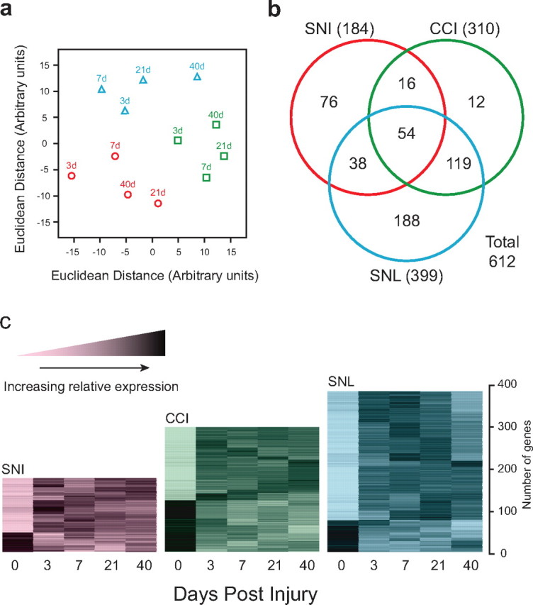
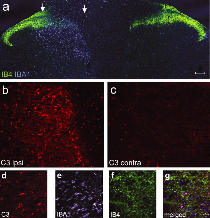
Microarray expression profiles reveal substantial changes in gene expression in the ipsilateral dorsal horn of the spinal cord in response to three peripheral nerve injury models of neuropathic pain. However, only 54 of the 612 regulated genes are commonly expressed across all the neuropathic pain models. Many of the commonly regulated transcripts are immune related and include the complement components C1q, C3, and C4, which we find are expressed only by microglia. C1q and C4 are, moreover, the most strongly regulated of all 612 regulated genes. In addition, we find that the terminal complement component C5 and the C5a receptor (C5aR) are upregulated in spinal microglia after peripheral nerve injury. Mice null for C5 had reduced neuropathic pain sensitivity, excluding C3a as a pain effector. C6-deficient rats, which cannot form the membrane attack complex, have a normal neuropathic pain phenotype. However, C5a applied intrathecally produces a dose-dependent, slow-onset cold pain in naive animals. Furthermore, a C5aR peptide antagonist reduces cold allodynia in neuropathic pain models. We conclude that induction of the complement cascade in spinal cord microglia after peripheral nerve injury contributes to neuropathic pain through the release and action of the C5a anaphylatoxin peptide.


Figures

Figure 1.
Figure 1.
Global expression profiles in neuropathic pain models. A, Multidimensional scaling display of the dissimilarities among the microarrays. Axes are in arbitrary units derived such that the distance between each pair of points in the XY plane is the most accurate possible representation of the Euclidean distance between the expression levels of all the genes measured on the corresponding pair of microarrays. SNI (red circles), CCI (green squares), and SNL (blue triangles) data are shown with time points as indicated. B, Venn diagram showing the number of regulated genes meeting fold difference and statistical thresholds in each model (SNI, CCI, or SNL). C, Temporal expression patterns of genes regulated in neuropathic pain models within the dorsal horn. Each gene was normalized according to mean 0, SD 1 and subjected to k-means clustering. The increased relative expression level is shown by increasing darkness.
Figure 2.
Figure 2.
A, Distribution of commonly regulated genes according to functional class. B, Genes related to immune function regulated in neuropathic pain models in the dorsal horn. Data shown for each gene are SNI (red circles), CCI (green squares), or SNL (blue triangles) after injury. Each plot is on a log base 2 scale, with the origin zero equivalent to onefold (nonregulation). The rat gene abbreviation, the maximum difference from the origin on the log base 2 scale, and conversion to linear scale are as indicated beneath each plot. Genes are sorted according to peak upregulation. C1qb, C1qg, C3, and C4 complement components; Aif1, Allograft inflammatory factor 1 (Iba1); Ctss, cathepsin S; Ctsh, cathepsin H; CCR5, C-C chemokine receptor 5; Ifngr, interferon-γ receptor 1; Cx3cr1, chemokine (C-X3-C motif) receptor 1.
Figure 3.
Figure 3.
A, Complement cascade and major effector mechanisms. B, Expression by in situ hybridization of the complement genes C1qb, C4, and C3 in naive spinal cord and spinal cord 3, 7, and 40 d after SNI. Inset, The number is fold difference from the microarrays. Scale bar, 100 μm.
Figure 4.
Figure 4.
Complement gene expression in microglia. Fluorescent in situ hybridization for C1q, C4, and C3 in the ipsilateral dorsal horn is shown. Each mRNA signal colocalizes with Iba1, a microglial marker, but not with NeuN (neuronal) or GFAP (astrocyte) markers. Staining of C1q was performed 3 d after injury, and staining of C4 and C3 was performed 7 d after injury. Scale bar, 15 μm.
Figure 5.
Figure 5.
A, Confocal microscopic photomontage of the lumbar dorsal horn of the rat spinal cord 5 d after SNI labeled with the c-fiber central terminal marker IB4 (green) and the microglial marker Iba1 (blue). The region between the arrows has reduced IB4 staining marking the central termination zone of the injured axons. B, C, Complement component C3 immunostaining in the ipsilateral (B) and contralateral (C) dorsal horn 5 d after SNI from the same section acquired using the same microsope settings. D–F, C3 immunoreactivity (5 d SNI; D) colocalizes with Iba1 (microglia; E) but not IB4 (c-fiber; F) staining. G, Overlay of images D–F. Scale bars: A–C, 100 μm; E, F, 10 μm.
Figure 6.
Figure 6.
A, C5aR immunoreactivity within the ipsilateral dorsal horn of the spinal cord 3 d after SNI colocalizes with Iba1 (first 3 panels), confirming localization to microglia. Scale bar, 10 μm. B, C5aR immunoreactivity in the ipsilateral L4–L5 lumbar dorsal horn increases from a low expression in naive animals peaking 3 d after SNI. Scale bar, 100 μm. C, High-power image of the naive dorsal horn section in B showing baseline C5aR expression. D, E, Change in C5aR mRNA (D) and C5 mRNA (E) in the dorsal horn of the spinal cord of rats after SNI relative to levels in uninjured animals, detected by quantitative real-time PCR (n = 4; ANOVA, *p < 0.05).
Figure 7.
Figure 7.
Induction of C3 in microglial cultures. A–D, Neonatal mouse microglial cell cultures under phase (A, C) and labeled with two microglial markers, Iba1 (B) and CD11b (D). E, Quantitative real-time PCR shows strong upregulation of C3 mRNA 24 h after treatment with LPS (10 μg/ml) and TNF-α (10 ng/ml) but not IL-1β (10 ng/ml). E–H, Complement C3 protein is upregulated in the presence of LPS (10 μg/ml; G) and TNF-α (10 ng/ml; H) compared with control (F) after 24 h. DAPI staining for nuclei (blue) is shown in all merged images. Scale bar, 50 μm. *p < 0.05 (t test).
Figure 8.
Figure 8.
C5a function. A, Paw withdrawal in response to a cold stimulus as a function of time subsequent to injection of C5a peptide into the intrathecal space of rats. Data for saline injection (filled squares; n = 7), 10 ng of C5a peptide (open circles; n = 7), and 100 ng of C5a peptide (open squares; n = 5) are shown. The area under the response-time curve (AUC) was calculated for each dose. There was a significant difference among the three doses by one-way ANOVA (F (2,16) = 8.72; p = 0.0027), with post hoc testing (Tukey's method) showing a significant difference between saline and 100 ng (p = 0.002) but not between 10 and 100 ng (p = 0.13) or between saline and 10 ng (p = 0.09). All data are mean ± SEM. B, Paw withdrawal in response to a cold stimulus (acetone evaporation) as a function of time subsequent to SNI injury in rats during continuous infusion of a C5a antagonist into the intrathecal space via an osmotic pump. Day −1 behavioral testing was before pump placement; day 0 behavioral testing was before SNI injury. There was a significant difference in the area under the response-time curve between C5a antagonist-treated (open squares; n = 12) and vehicle-treated (filled squares; n = 11) animals by Welch's two-sample t test (t = 2.17; df = 20.4; p = 0.042).

References

    1. Abbadie C, Lindia JA, Cumiskey AM, Peterson LB, Mudgett JS, Bayne EK, DeMartino JA, MacIntyre DE, Forrest MJ. Impaired neuropathic pain responses in mice lacking the chemokine receptor CCR2. Proc Natl Acad Sci USA. 2003;100:7947–7952. - PMC - PubMed
    1. Albrecht EA, Chinnaiyan AM, Varambally S, Kumar-Sinha C, Barrette TR, Sarma JV, Ward PA. C5a-induced gene expression in human umbilical vein endothelial cells. Am J Pathol. 2004;164:849–859. - PMC - PubMed
    1. Arruda JL, Colburn RW, Rickman AJ, Rutkowski MD, DeLeo JA. Increase of interleukin-6 mRNA in the spinal cord following peripheral nerve injury in the rat: potential role of IL-6 in neuropathic pain. Brain Res Mol Brain Res. 1998;62:228–235. - PubMed
    1. Bennett GJ, Xie YK. A peripheral mononeuropathy in rat that produces disorders of pain sensation like those seen in man. Pain. 1988;33:87–107. - PubMed
    1. Blackshaw S, Snyder SH. Parapinopsin, a novel catfish opsin localized to the parapineal organ, defines a new gene family. J Neurosci. 1997;17:8083–8092. - PMC - PubMed

Publication types

MeSH terms