Three-dimensional mapping of hippocampal anatomy in unmedicated and lithium-treated patients with bipolar disorder
- PMID: 17687266
- PMCID: PMC6693586
- DOI: 10.1038/sj.npp.1301507
Three-dimensional mapping of hippocampal anatomy in unmedicated and lithium-treated patients with bipolar disorder
Abstract
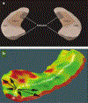
Declarative memory impairments are common in patients with bipolar illness, suggesting underlying hippocampal pathology. However, hippocampal volume deficits are rarely observed in bipolar disorder. Here we used surface-based anatomic mapping to examine hippocampal anatomy in bipolar patients treated with lithium relative to matched control subjects and unmedicated patients with bipolar disorder. High-resolution brain magnetic resonance images were acquired from 33 patients with bipolar disorder (21 treated with lithium and 12 unmedicated), and 62 demographically matched healthy control subjects. Three-dimensional parametric mesh models were created from manual tracings of the hippocampal formation. Total hippocampal volume was significantly larger in lithium-treated bipolar patients compared with healthy controls (by 10.3%; p=0.001) and unmedicated bipolar patients (by 13.9%; p=0.003). Statistical mapping results, confirmed by permutation testing, revealed localized deficits in the right hippocampus, in regions corresponding primarily to cornu ammonis 1 subfields, in unmedicated bipolar patients, as compared to both normal controls (p=0.01), and in lithium-treated bipolar patients (p=0.03). These findings demonstrate the sensitivity of these anatomic mapping methods for detecting subtle alterations in hippocampal structure in bipolar disorder. The observed reduction in subregions of the hippocampus in unmedicated bipolar patients suggests a possible neural correlate for memory deficits frequently reported in this illness. Moreover, increased hippocampal volume in lithium-treated bipolar patients may reflect postulated neurotrophic effects of this agent, a possibility warranting further study in longitudinal investigations.
Conflict of interest statement
DISCLOSURES
The authors have no conflicts of interest and no relevant financial disclosures to declare.
Figures


References
-
- Altshuler LL, Ventura J, van Gorp WG, Green MF, Theberge DC, Mintz J (2004). Neurocognitive function in clinically stable men with bipolar I disorder or schizophrenia and normal control subjects. Biol Psychiatry 56: 560–569. - PubMed
-
- Apostolova LG, Dutton RA, Dinov ID, Hayashi KM, Toga AW, Cummings JL et al. (2006). Conversion of mild cognitive impairment to Alzheimer disease predicted by hippocampal atrophy maps. Arch Neurol 63: 693–699. - PubMed
-
- Bearden CE, Glahn DC, Monkul ES, Barrett J, Najt P, Kaur S et al. (2006). Sources of declarative memory impairment in bipolar disorder: mnemonic processes and clinical features. J Psychiatr Res 40: 47–58. - PubMed
-
- Bech P, Bolwig TG, Kramp P, Rafaelsen OJ (1979). The Bech–Rafaelsen mania scale and the hamilton depression scale. Acta Psychiatr Scand 59: 420–430. - PubMed
Publication types
MeSH terms
Substances
Grants and funding
- K23 MH074644/MH/NIMH NIH HHS/United States
- P30 MH030915/MH/NIMH NIH HHS/United States
- AG016570/AG/NIA NIH HHS/United States
- R21 RR019771/RR/NCRR NIH HHS/United States
- K24 RR020571/RR/NCRR NIH HHS/United States
- R01 HD050735/HD/NICHD NIH HHS/United States
- K23 MH001736/MH/NIMH NIH HHS/United States
- K23 MH074644-01/MH/NIMH NIH HHS/United States
- MH 30915/MH/NIMH NIH HHS/United States
- RR020571/RR/NCRR NIH HHS/United States
- MH 01736/MH/NIMH NIH HHS/United States
- HD050735/HD/NICHD NIH HHS/United States
- EB01651/EB/NIBIB NIH HHS/United States
- RR019771/RR/NCRR NIH HHS/United States
- P50 AG016570/AG/NIA NIH HHS/United States

