. 2007 Summer;12(2):132-41.
doi: 10.1379/csc-233r.1.

Heat stress inhibits skeletal muscle hypertrophy

Affiliations

Heat stress inhibits skeletal muscle hypertrophy

Bruce C Frier et al. Cell Stress Chaperones. 2007 Summer.

Abstract

Heat shock proteins (Hsps) are molecular chaperones that aid in protein synthesis and trafficking and have been shown to protect cells/tissues from various protein damaging stressors. To determine the extent to which a single heat stress and the concurrent accumulation of Hsps influences the early events of skeletal muscle hypertrophy, Sprague-Dawley rats were heat stressed (42 degrees C, 15 minutes) 24 hours prior to overloading 1 plantaris muscle by surgical removal of the gastrocnemius muscle. The contralateral plantaris muscles served as controls. Heat-stressed and/or overloaded plantaris muscles were assessed for muscle mass, total muscle protein, muscle protein concentration, Type I myosin heavy chain (Type I MHC) content, as well as Hsp72 and Hsp25 content over the course of 7 days following removal of the gastrocnemius muscle. As expected, in non-heat-stressed animals, muscle mass, total muscle protein and MHC I content were significantly increased (P < 0.05) following overload. In addition, Hsp25 and Hsp72 increased significantly after 2 and 3 days of overload, respectively. A prior heat stress-elevated Hsp25 content to levels similar to those measured following overload alone, but heat stress-induced Hsp72 content was increased significantly greater than was elicited by overload alone. Moreover, overloaded muscles from animals that experienced a prior heat stress showed a lower muscle mass increase at 5 and 7 days; a reduced total muscle protein elevation at 3, 5, and 7 days; reduced protein concentration; and a diminished Type I MHC content accumulation at 3, 5, and 7 days relative to nonheat-stressed animals. These data suggest that a prior heat stress and/or the consequent accumulation of Hsps may inhibit increases in muscle mass, total muscle protein content, and Type I MHC in muscles undergoing hypertrophy.


Figures
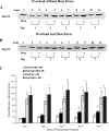
Fig 1.
Fig 1.
Hsp72 content in plantaris muscles following heat stress and overload. (A) Representative blot of Hsp72 content in non–heat-stressed animals. Lane 1: Pure Hsp72 (2ng); lane 2: contralateral control plantaris (−) day 1; lane 3: overloaded plantaris (+) day 1; lane 4: contralateral control plantaris (−) day 2; lane 5: overloaded plantaris (+) day 2; lane 6: contralateral control plantaris (−) day 3; lane 7: overloaded plantaris (+) day 3; lane 8: contralateral control plantaris (−) day 5; lane 9: overloaded plantaris (+) day 5; lane 10: contralateral control plantaris (−) day 7; lane 11: overloaded plantaris (+) day 7. Hsp72 content was significantly increased (P < 0.05) in the overloaded plantaris muscle on days 3, 5, and 7. (B) Representative blot of Hsp72 content in heat-stressed animals. Lanes are as described for (A). Hsp72 content in the plantaris muscle is not significantly different between contralateral control plantaris (−) and overloaded plantaris (+) at any time point. (C) Hsp72 content in all plantaris muscles on days 1, 3, 5, and 7. Data are expressed as mean ± SEM. *Significantly different (P < 0.05) from non–heat-stressed controls and non–heat-stressed overloaded plantaris muscles. †Significantly different (P < 0.05) from non–heat-stressed contralateral control plantaris
Fig 2.
Fig 2.
Hsp25 content in plantaris muscles following heat stress and overload. (A) Western blot of Hsp25 accumulation in non–heat-stressed animals. Lane 1: Pure Hsp25 (4 ng); lane 2: contralateral control plantaris (−) day 1; lane 3: overloaded plantaris (+) day 1; lane 4: contralateral control plantaris (−) day 2; lane 5: overloaded plantaris (+) day 2; lane 6: contralateral control plantaris (−) day 3; lane 7: overloaded plantaris (+) day 3; lane 8: contralateral control plantaris (−) day 5; lane 9: overloaded plantaris (+) day 5; lane 10: contralateral control plantaris (−) day 7; lane 11: overloaded plantaris (+) day 7. Hsp25 content is significantly increased (P < 0.05) in the non-heat stressed overloaded (+) plantaris muscle on days 3, 5, and 7. (B) Western blot of Hsp25 content in heat stressed animals. Lane 1: contralateral control plantaris (−) day 1; lane 2: overloaded plantaris (+) day 1; lane 3: contralateral control plantaris (−) day 2; lane 4: overloaded plantaris (+) day 2; lane 5: contralateral control plantaris (−) day 3; lane 6: overloaded plantaris (+) day 3; lane 7: contralateral control plantaris (−) day 5; lane 8: overloaded plantaris (+) day 5; lane 9: contralateral control plantaris (−) day 7; lane 10: overloaded plantaris (+) day 7; lane 11: pure Hsp25 (4 ng). Hsp25 content in overloaded heat-stressed plantaris (+) muscles is significantly elevated (P < 0.05) from the heat-stressed contralateral control (−) plantaris beginning on day 2. (C) Composite data representing Hsp25 content in all muscles. Data are expressed as mean ± SEM. *Significantly different (P < 0.05) from non–heat-stressed control muscles
Fig 3.
Fig 3.
Plantaris muscle protein content is increased following overload. Values are means ± SEM for total protein (mg). *Significantly different (P < 0.05) from contralateral controls plantaris. †Significantly different (P < 0.05) from all other tissues
Fig 4.
Fig 4.
Protein concentration comparison between non–heat-stressed and heat-stressed muscles. Values are means ± SEM for protein concentration (μg/mg). *Significantly different (P < 0.05) from heat-stressed plantaris muscles
Fig 5.
Fig 5.
Plantaris Type I MHC content is decreased following heat stress. Values are means ± SEM for total protein (mg). *Significantly different (P < 0.05) from all other tissues. †Significantly different (P < 0.05) from contralateral control plantaris

References

    1. Bischoff R 1994 The satellite cell and muscle regeneration. In: Myogenesis, ed Engel AG, Franszini-Armstrong C. McGraw-Hill, New York, 97–118.
    1. Currie RW, Tanguay RM, Kingma JG Jr.. Heat-shock response and limitation of tissue necrosis during occlusion/reperfusion in rabbit hearts. Circulation. 1993;87:963–971.0009-7322(1993)087[0963:HRALOT]2.0.CO;2 - PubMed
    1. Halevy O, Krispin A, Leshem Y, McMurtry JP, Yahav S. Early-age heat exposure affects skeletal muscle satellite cell proliferation and differentiation in chicks. Am J Physiol Regul Integr Comp Physiol. 2001;281:R302–R309.1522-1490(2001)281[R302:EHEASM]2.0.CO;2 - PubMed
    1. Hubbard RW, Ianuzzo CD, Mathew WT, Linduska JD. Compensatory adaptations of skeletal muscle composition to a long-term functional overload. Growth. 1975;39:85–93.0258-7173(1975)039[0085:CAOSMC]2.0.CO;2 - PubMed
    1. Ianuzzo CD, Chen V. Metabolic character of hypertrophied rat muscle. J Appl Physiol. 1979;46:738–742.8750-7587(1979)046[0738:MCOHRM]2.0.CO;2 - PubMed

Publication types

LinkOut - more resources