. 2007 Aug 9:4:57.
doi: 10.1186/1742-4690-4-57.

Endogenous TGF-beta activation by reactive oxygen species is key to Foxp3 induction in TCR-stimulated and HIV-1-infected human CD4+CD25- T cells

Affiliations

Endogenous TGF-beta activation by reactive oxygen species is key to Foxp3 induction in TCR-stimulated and HIV-1-infected human CD4+CD25- T cells

Shoba Amarnath et al. Retrovirology. .

Abstract

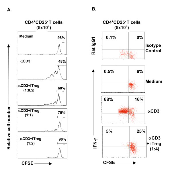
Background: CD4+CD25+ T regulatory cells (Tregs) play an important role in regulating immune responses, and in influencing human immune diseases such as HIV infection. It has been shown that human CD4+CD25+ Tregs can be induced in vitro by TCR stimulation of CD4+CD25- T cells. However, the mechanism remains elusive, and intriguingly, similar treatment of murine CD4+CD25- cells did not induce CD4+CD25+Foxp3+ Tregs unless exogenous TGF-beta was added during stimulation. Thus, we investigated the possible role of TGF-beta in the induction of human Tregs by TCR engagement. We also explored the effects of TGF-beta on HIV-1 infection mediated induction of human Tregs since recent evidence has suggested that HIV-1 infection may also impact the generation of Tregs in infected patients.

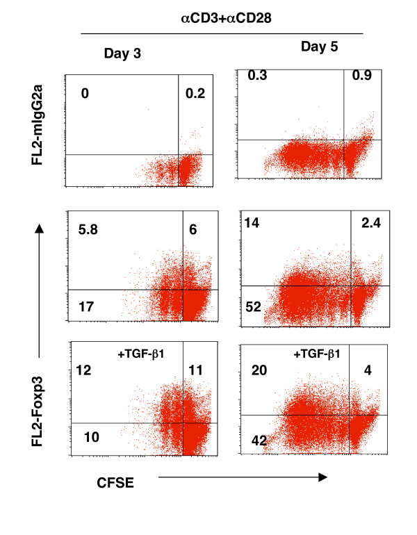
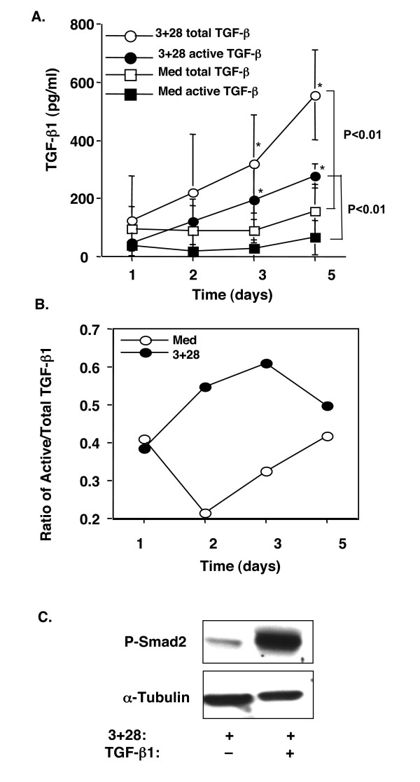
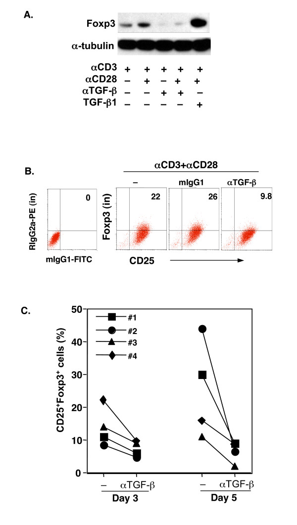
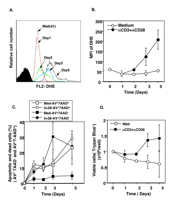
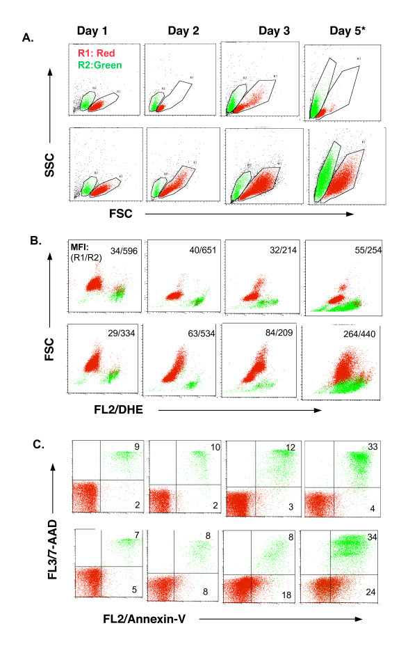
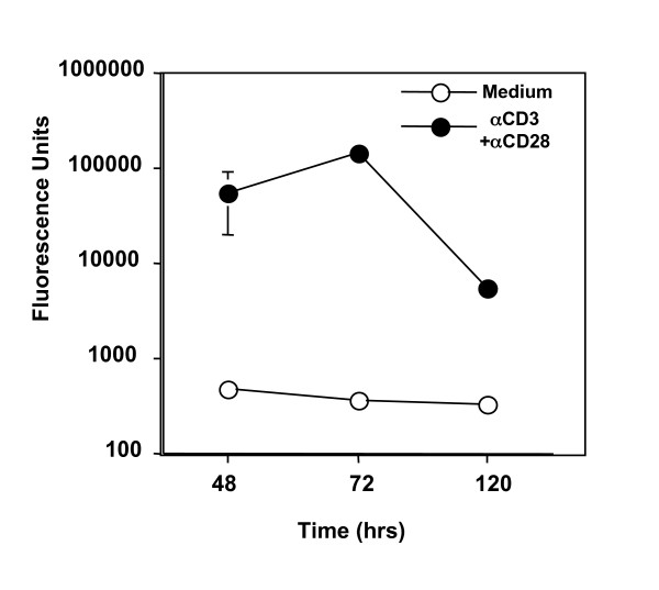
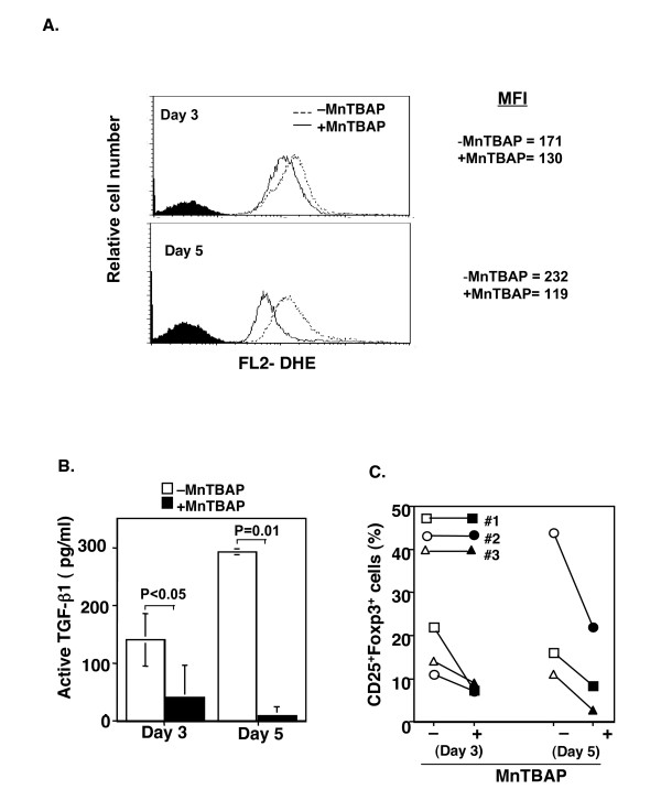
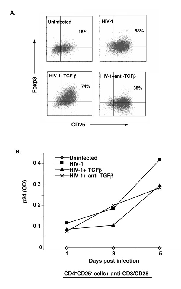
Results: We show here that endogenous TGF-beta is key to TCR induction of Foxp3 in human CD4+CD25- T cells. These events involve, first, the production of TGF-beta by TCR and CD28 stimulation and the activation of latent TGF-beta by reactive oxygen species generated from the activated T cells. Biologically active TGF-beta then engages in the induction of Foxp3. Neutralization of active TGF-beta with anti-TGF-beta antibody or elimination of ROS with MnTBAP abrogated Foxp3 expression. HIV-1 infection enhanced Foxp3 expression in activated CD4+CD25- T cells; which was also abrogated by blockade of endogenous TGF-beta.

Conclusion: Several conclusions can be drawn from this work: (1) TCR and CD28-induced Foxp3 expression is a late event following TCR stimulation; (2) TGF-beta serves as a link in Foxp3 induction in human CD4+CD25- T cells following TCR stimulation, which induces not only latent, but also active TGF-beta; (3) the activation of TGF-beta requires reactive oxygen species; (4) HIV infection results in an increase in Foxp3 expression in TCR-activated CD25- T cells, which is also associated with TGF-beta. Taken together, our findings reinforce a definitive role of TGF-beta not only in the generation of Tregs with respect to normal immune responses, but also is critical in immune diseases such as HIV-1 infection.


Figures

Figure 1
Figure 1
TCR stimulation of human CD4+CD25- T cells induces Foxp3. Highly purified CD4+CD25- T cells (98–99%) were stimulated with the indicated regimen in X-Vivo 20 serum-free medium, and Foxp3 mRNA and protein were examined. A. Cells were cultured for 48–72 hours. RNA was isolated and cDNA synthesized for assessing the expression of Foxp3 by real-time PCR. Freshly isolated CD4+CD25+ T cells were used as a positive control for Foxp3 expression. Values are expressed as the normalized ratio of Foxp3 to GAPDH. B. Analysis of Foxp3 protein with Western blot. The experiments were repeated three times with similar results. C-E. Analysis of intracellular Foxp3 protein at the single-cell level by FACS. Freshly isolated CD4+CD25- cells (Fresh) or cultured cells at the indicated time were stained with FITC-anti-CD25 (surface) and PE-anti-Foxp3 (intracellular) and analyzed on FACScalibur. A representative FACS profile is shown as dot plots of CD25 versus Foxp3 (C). The quadrant gates were set according to the negative isotype control antibodies in the respective cells. The kinetics of the percentage (D) and total number (E) of CD25+Foxp3+cells are shown as Mean ± SD of each group at each time point (n = 3 to 6). Med: Medium; αCD3: anti-CD3 mAb; αCD28: anti-CD28 mAb. * indicates a different donor.
Figure 2
Figure 2
Foxp3+ T cells exist in both non-proliferating (CFSE+hi) and dividing (CFSE+low) TCR-stimulated CD4+CD25- T cells. CD4+CD25- T cells were labeled with CFSE (2.5 μM) and cultured with anti-CD3 and anti-CD28 for 3 and 5 days. Cells were then counter-stained intracellularly with PE-conjugated anti-Foxp3 antibody. The cells were analyzed with FACS and a representative profile of CFSE vs. Foxp3 or its control antibody (mIgG2a) is displayed. The experiments were repeated three times with similar results. Data not shown here are the cultures with cells in medium alone. No CFSE dilution (CFSE+low) or Foxp3+ cells were observed.
Figure 3
Figure 3
TCR induced CD25+Foxp3+ T cells were immunosuppressive to CD4+CD25- T cell proliferation in vitro. A. CD4+CD25- T cells were cultured with anti-CD3 and anti-CD28 for 5 days. The converted CD4+CD25+Foxp3+ T cells were purified and washed extensively. The converted Tregs were then used at varying concentrations in a co-culture suppression assay along with CD4+CD25- (5 × 10⁴) T cells pre-labeled with CFSE as responders and autologous monocytes (2 × 10⁵) as accessory cells. Anti-CD3 antibody was added into the start of the co-culture suppression assay (0.5 μg/ml). CFSE dilution of responder cells was measured after 72 hrs using flow cytometry. B. IFN-γ production of responder cells in the co-culture assay as detected by flow cytometry after 72 hrs. iTreg: induced Foxp3+CD4+CD25+ T cells. The experiment was repeated for three times with similar results.
Figure 4
Figure 4
TCR and CD28 stimulation of human CD4+CD25- T cells produced TGF-β and exhibited phosphorylation of Smad2. A. CD4+CD25- T cells (1 × 10⁶/ml) were cultured with anti-CD3 and anti-CD28 in X-Vivo 20 serum-free medium for the indicated time points. Cell-free supernatants were either untreated (for active TGF-β) or treated with 1 N HCl (for total TGF-β) followed by ELISA for TGF-β1 measurement. The values are shown as Mean ± SD of individuals in each group at each time point (n = 3 to 9). B. The relative ratio of active to total TGF-β is shown in each time point as in A. C. Western blot analysis of P-Smad2 in cultured CD4+CD25- T cells (72 hrs). Whole cell lysis protein (70 μg/ml) was loaded into each lane. P-Smad2 was detected with anti-P-Smad2 antibody. α-tubulin was used as host protein control. 3+28: anti-CD3+anti-CD28; Med: medium.
Figure 5
Figure 5
Neutralization of endogenous TGF-β abrogated TCR-induced Foxp3 expression. A. Western blot analysis of Foxp3 protein in cultured CD4+CD25- T cells with indicated reagents (72 hrs). B. FACS analysis of intracellular Foxp3 protein cultured with TCR and CD28 in the presence of anti-TGF-β1,2,3 (αTGF-β) or control (mIgG1) antibodies (72 hr). The data are shown for a representative donor. The values are presented as the percentage of CD25+Foxp3+ T cells. C. CD25+Foxp3+ T cells (%) in the TCR- and CD28-stimulated CD4+CD25- T cells in the absence (-) or presence of anti-TGF-β1,2,3 antibody (αTGF-β) at days 3 and 5. Each symbol represents one donor.
Figure 6
Figure 6
TCR and CD28 stimulation induced ROS production and increased T cell apoptosis. CD4+CD25- T cells were stimulated with anti-CD3 and anti-CD28 antibodies for the indicated time points and then intracellular ROS was stained with DHE. The mean fluorescence intensity (MFI) of DHE in a single cell was measured with FACS. A aliquot of cells was stained with Annexin-V and 7-AAD to analyze the early apoptotic (Annexin-V+7-AAD-) and late apoptotic/dead (Annexin+7-AAD+) cells. The cells from each cultured well were also examined for viable cells by trypan blue exclusion assay. A. A representative histogram profile of DHE staining on the different days. The filled histogram is the un-labeled cells (negative control). B. The values are displayed as the Mean ± SD of the MFI of DHE between stimulated (αCD3+αCD28) and non-stimulated (medium) live T cells (R1 gated cells in Fig. S1) at the indicated time points (n = 2 to 5). C. The values are presented as the Mean ± SD of the early apoptotic (Annexin+7-AAD-) and dead/late apoptotic (Annexin+7-AAD+) between anti-CD3 and anti-CD28 (3+28) and non-stimulated (medium) cells (n = 2 to 5). D. The values are shown as the Mean ± SD of the live cells (trypan blue negative) per well. The original cell number was 1 × 10⁶ per well(24-well plate; n = 2 to 5).
Figure 7
Figure 7
A representative FACS profile of cell size (A), DHE fluorescence (B) and apoptotic cells (C) between anti-CD3 plus anti-CD28-stimulated and medium-treated CD4+CD25- T cells is displayed. A. Profile of FSC vs SSC is displayed to show the cell size. The cells were electronically gated as two populations based on their size. R1 (red) represents live or early apoptotic cells (see C). R2 (green) represents dead and/or late apoptotic cells (see C). B. Profile of DHE fluorescence (ROS+) on FL-2 vs. FSC of R1 and R2 cells. The values are shown as the MFI of R1 and R2 cells (R1/R2). Data not shown here are the MFI of unlabeled cells (negative control for DHE staining) on FL2, which is usually < 10. C. The profile of Annexin-V vs. 7-AAD staining of cultured cells compensating the R1 and R2 regions as gated in A. The quadrant gates were set according to the negative isotype control antibodies in the respective cells.
Figure 8
Figure 8
The cell-free supernatant from TCR stimulated CD4+CD25- T cell culture contained ROS. CD4+CD25- T cells were cultured with anti-CD3 and anti-CD28 for the indicated time points and ROS in the culture supernatant was detected using DCFH-DA as described in the Method section. Oxidation of DCFH-DA was measured using a spectrofluorometer at wavelength 485/535 nm and is represented as fluorescent units. The experiment was repeated twice with similar results.
Figure 9
Figure 9
Neutralization of ROS with MnTBAP abrogated active TGF-β and reduced CD25+Foxp3+ T cells. CD4+CD25- T cells were cultured with anti-CD3 and anti-CD28 antibodies in the presence or absence of MnTBAP (100 μM) for 3 and 5 days. The intracellular ROS production was determined by DHE staining. Active TGF-β in the supernatants was determined with ELISA. The intracellular Foxp3 protein was determined by FACS staining. A. A representative overlay of histograms of ROS in the cultured T cells with (+MnTBAP) and without (-MnTBAP) MnTBAP at days 3 and 5. The filled histograms were from unlabeled cells as negative control for DHE staining. The experiment was repeated three times with similar results. B. The values are shown as the Mean ± SD of active TGF-β1 in the culture supernatants at days 3 and 5 (n = 2). C. MnTBAP reduced CD25+Foxp3+ T cells. Each symbol represents one individual.
Figure 10
Figure 10
HIV infection upregulated Foxp3 expression in TCR activated CD4+CD25- T cells. A. Purified CD4+CD25- T cells were infected with HIV1 (HIV NLA-3) and cultured with anti-CD3 and anti-CD28 antibodies in the absence (HIV-1) or presence of active TGF-β1 (2 ng/ml) (HIV-1+TGF-β) or anti-TGF-β1,2,3 antibody (HIV-1+anti-TGF-β) for 3 (data not shown) and 5 days. T cells were then stained for surface CD25 and intracellular Foxp3. A representative of two experiments is displayed. A parallel culture of TCR- and CD28-stimulated CD25- T cells without HIV infection (uninfected) was used as control. B. The supernatants from the same cultures in A were collected at the indicated time points and tested for HIV p24 with ELISA. The experiment was repeated twice with similar results.

References

    1. Fontenot JD, Rasmussen JP, Williams LM, Dooley JL, Farr AG, Rudensky AY. Regulatory T cell lineage specification by the forkhead transcription factor foxp3. Immunity. 2005;22:329–341. doi: 10.1016/j.immuni.2005.01.016. - DOI - PubMed
    1. Kuniyasu Y, Takahashi T, Itoh M, Shimizu J, Toda G, Sakaguchi S. Naturally anergic and suppressive CD25(+)CD4(+) T cells as a functionally and phenotypically distinct immunoregulatory T cell subpopulation. Int Immunol. 2000;12:1145–1155. doi: 10.1093/intimm/12.8.1145. - DOI - PubMed
    1. Shevach EM. CD4+ CD25+ suppressor T cells: more questions than answers. Nat Rev Immunol. 2002;2:389–400. - PubMed
    1. Bluestone JA, Abbas AK. Natural versus adaptive regulatory T cells. Nat Rev Immunol. 2003;3:253–257. doi: 10.1038/nri1032. - DOI - PubMed
    1. Powrie F, Maloy KJ. Immunology. Regulating the regulators. Science. 2003;299:1030–1031. doi: 10.1126/science.1082031. - DOI - PubMed

Publication types

MeSH terms

Substances