. 2007 Nov 25;368(2):331-41.
doi: 10.1016/j.virol.2007.06.042. Epub 2007 Aug 3.

Structural basis for the binding of the neutralizing antibody, 7D11, to the poxvirus L1 protein

Affiliations

Structural basis for the binding of the neutralizing antibody, 7D11, to the poxvirus L1 protein

Hua-Poo Su et al. Virology. .

Abstract

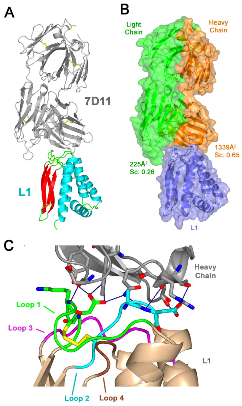
Medical countermeasures to prevent or treat smallpox are needed due to the potential use of poxviruses as biological weapons. Safety concerns with the currently available smallpox vaccine indicate a need for research on alternative poxvirus vaccine strategies. Molecular vaccines involving the use of proteins and/or genes and recombinant antibodies are among the strategies under current investigation. The poxvirus L1 protein, encoded by the L1R open reading frame, is the target of neutralizing antibodies and has been successfully used as a component of both protein subunit and DNA vaccines. L1-specific monoclonal antibodies (e.g., mouse monoclonal antibody mAb-7D11, mAb-10F5) with potent neutralizing activity bind L1 in a conformation-specific manner. This suggests that proper folding of the L1 protein used in molecular vaccines will affect the production of neutralizing antibodies and protection. Here, we co-crystallized the Fab fragment of mAb-7D11 with the L1 protein. The crystal structure of the complex between Fab-7D11 and L1 reveals the basis for the conformation-specific binding as recognition of a discontinuous epitope containing two loops that are held together by a disulfide bond. The structure of this important conformational epitope of L1 will contribute to the development of molecular poxvirus vaccines and also provides a novel target for anti-poxvirus drugs. In addition, the sequence and structure of Fab-7D11 will contribute to the development of L1-targeted immunotherapeutics.


Figures

Fig. 1
Fig. 1
Interaction of mAb-7D11, F(ab′)2, and Fab fragments with purified VACV and recombinant L1. (A) Binding to purified VACV. Serial twofold dilutions of mAb-7D11, 7D11-F(ab′)2, and 7D11-Fab or negative control antibody mAb-3D7 starting at 80 μg/ml were tested for the capacity to bind purified virus by VACV-infected-cell lysate ELISA. Some antigen-coated plates were treated with UV light (grey dashed lines) before incubation with mAb-7D11 (MAb-7D11 UV). Each symbol represents the mean ± standard deviation (SD) of duplicate samples. (B) Binding to recombinant L1. Serial twofold dilutions of mAb-7D11, 7D11-F(ab′)2, and 7D11-Fab or negative control antibody mAb-10F10 (anti-A33) starting at 80 μg/ml were tested for the capacity to bind recombinant L1 by ELISA. Purified L1 at 100 ng/well was untreated or treated with UV light (grey dashed lines). Each symbol represents the mean ± SD of duplicate samples. The x-axes in A and B are plotted on log(2) scales. The smallest error bars in A and B are hidden beneath the symbols and are not seen.
Fig. 2
Fig. 2
Plaque reduction-neutralization test using mAb-7D11, 7D11-F(ab′)2, and 7D11-Fab. (A) VACV was incubated with purified mAb-7D11, 7D11-F(ab′)2, or 7D11-Fab at 37°C for 1 h. Virus-antibody mixtures or untreated virus were adsorbed on to confluent monolayers of BSC-1 cells, followed the addition of a semi-solid overlay of 1.5% methylcellulose as described in Methods. After 4 d, plaques were counted after staining with 3% crystal violet. The percent neutralization of plaque formation by antibody addition relative to untreated virus is plotted versus concentration on a log(2) scale. (B) VACV was incubated with mAb-7D11 or 7D11-Fab at the concentrations indicated, for 1 h at 37°C. One sample, incubated with Fab, was then incubated with anti-Fab antibodies (1:100) for 1 h. The other samples were incubated for an additional 1 h in the absence of anti-Fab antibodies. After incubation, the virus was added to BSC-1 cells and plaque formation analyzed as described in A. (C) Confluent monolayers of BSC-1 cells were chilled on ice and incubated with ~50 PFU of VACV strain IHD-J per well for 1 h at 4°C. After adsorption, virus was removed and mAb-7D11 or 7D11-Fab was added to the wells at the indicated concentrations for 1 h at 4°C. After incubation, anti-Fab antibodies (1:100) were added to the wells of some samples for another 1 h at 4°C. Plates were then incubated at 37°C and plaques were visualized as described in A.
Fig. 3
Fig. 3
Structure of 7D11-Fab bound to L1. (A) 7D11-Fab recognizes the loops connecting the helices to the β strands of L1. The ribbon diagram shows L1 bound by the 7D11-Fab (gray). L1 is colored by secondary structure with β sheets in red, helices in blue, and loops in green. Disulfide bonds are shown in yellow. (B) The heavy chain of 7D11-Fab provides most of the interactions with L1. In the same orientation as in A, the surface diagram is colored by polypeptide chains with the 7D11 heavy chain in orange, the light chain in green, and L1 in blue. The calculated total buried surface area and the shape complementarity statistic between L1 and each of the antibody chains are indicated. (C) 7D11-Fab makes numerous hydrogen bonds and van der Waals interactions with loops 1 and 2 of L1. Oriented as in parts A and B, and numbered from N-terminus to C-terminus, the four loops of L1 are colored as follows: loop 1 (green), loop 2 (blue), loop 3 (purple), and loop 4 (brown). These loops are held together by a disulfide bond (yellow) between Cys 34 in loop 1 and Cys 57 in loop 2. The carbon atoms are colored based on the loop that the residue is in, nitrogen atoms are blue, and oxygen atoms are red. Hydrogen bonds displayed as dashed lines in blue show extensive bonding with loop 1 (green) and loop 2 (blue) of L1.
Fig. 4
Fig. 4
Interaction of anti-L1 monoclonal antibodies with mutated L1. (A) COS-7 cells were transfected with pWRG/TPA-L1R (black line), pWRG/TPA-L1R D35N (grey line), or empty pWRG vector (solid grey). Cells were incubated with 1:100 dilutions of mAb-7D11 (anti-L1), mAb-10F5 (anti-L1), or a control antibody mAb-10F10 (anti-A33). A anti-mouse secondary antibody (1:500) conjugated to Alexa 488 fluorochrome was added and cells were analyzed by flow cytometry using a FACSCalibur flow cytometer. For each sample, 10,000 cells were counted. (B) Hydrogen bonds involved in the binding by the 7D11-Fab to Asp35 of L1 as determined in the crystal structure are shown in blue. The atoms of critical residues are shown as sticks. The carbon atoms retain the color of the loops to which they belong: loop 1 (green), loop 2 (blue), loop 3 (purple), and loop 4 (brown). In the structure (upper panel), Asp35 (green) makes hydrogen bonds with the side chains of Trp33 and Tyr50 on the heavy chain (gray). Also shown is the hydrogen bond between the carbonyl oxygen of Ala59 on L1 (blue) and Nδ2 of Asn52 of the heavy chain (gray). In the lower panel, the predicted binding of mAb-7D11 to the D35N mutant of L1 (Model) is shown. The hydrogen bond between Asn35 of L1 (green) and Tyr50 of the heavy chain would be analogous to the bond made with Asp35. A new hydrogen bond (red) can be formed between Asn35 of L1 and an alternate rotamer of Asn52 of the Fab heavy chain, while maintaining the hydrogen bond (red) between Ala59 of L1 and Asn52 of the heavy chain. The L1:7D11-Fab structure has been rotated 180° around the vertical axis relative to its orientation in Fig. 3C. (C) Alignment of the L1 ectodomain sequences from VACV, Variola virus, and monkeypox virus in which residues are shown that differ from the vaccinia virus sequence. Residues of the four loops are shaded as in Fig. 3C: loop 1 (green), loop 2 (blue), loop 3 (purple), and loop 4 (brown). Residues involved in van der Waals interactions are marked with one dot and residues that also mediate hydrogen bonds are marked with two dots above the sequence. Cysteines are highlighted in yellow.

References

    1. Adams PD, Pannu NS, Read R, Brunger AT. Cross-validated maximum likelihood enhances crystallographic simulated annealing refinement. Proc Natl Acad Sci USA. 1997;94(10):5018–5023. - PMC - PubMed
    1. Aldaz-Carroll L, Whitbeck J, Ponce De Leon M, Lou H, Pannell L, Lebowitz J, Fogg C, White CL, Moss B, Cohen GH, Eisenberg R. Physical and immunological characterization of a recombinant secreted form of the membrane protein encoded by the vaccinia virus L1R gene. Virology. 2005;341(1):59–71. - PubMed
    1. Belongia E, Naleway A. Smallpox vaccine: the good, the bad, and the ugly. Clin Med Res. 2003;1(2):87–92. - PMC - PubMed
    1. Blouch R, Byrd C, Hruby D. Importance of disulphide bonds for vaccinia virus L1R protein function. Virol J. 2005;2:91. - PMC - PubMed
    1. Chen Z, Earl P, Americo J, Damon I, Smith SK, Zhou Y, Yu F, Sebrell A, Emerson S, Cohen G, Eisenberg R, Svitel J, Schuck P, Satterfield W, Moss B, Purcell R. Chimpanzee/human mAbs to vaccinia virus B5 protein neutralize vaccinia and smallpox viruses and protect mice against vaccinia virus. Proc Natl Acad Sci USA. 2006;103(6):1882–1887. - PMC - PubMed

Publication types

MeSH terms

Associated data