. 2007 Aug;142(2):170-9.
doi: 10.1016/j.surg.2007.04.015.

Norepinephrine suppresses wound macrophage phagocytic efficiency through alpha- and beta-adrenoreceptor dependent pathways

Affiliations

Norepinephrine suppresses wound macrophage phagocytic efficiency through alpha- and beta-adrenoreceptor dependent pathways

Ankush Gosain et al. Surgery. 2007 Aug.

Abstract

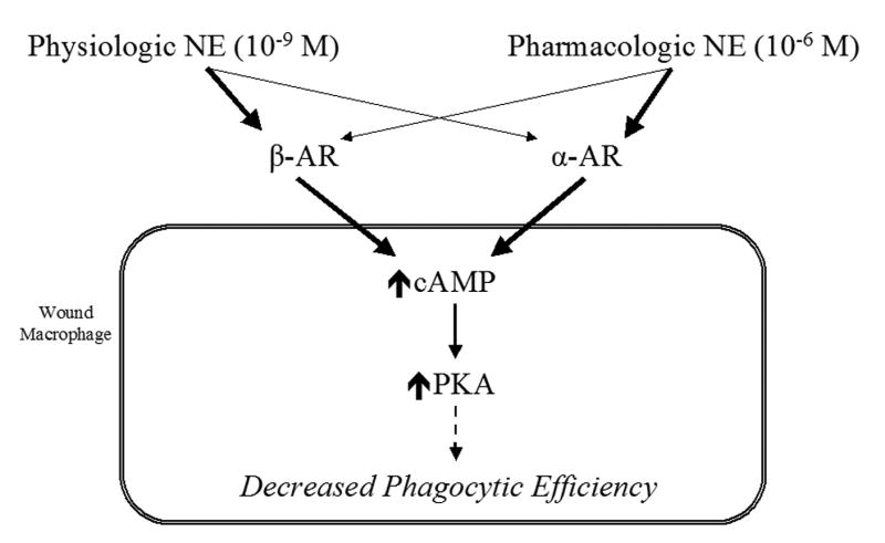
Background: The systemic response to injury is characterized by massive release of norepinephrine (NE) into the circulation as a result of global sympathetic activation. We have recently demonstrated that NE modulates the recruitment of macrophages to the cutaneous wound. We hypothesized that NE suppresses wound macrophage phagocytic function through canonical adrenergic signaling pathways.

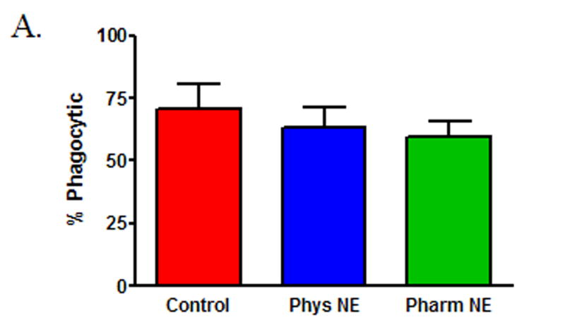
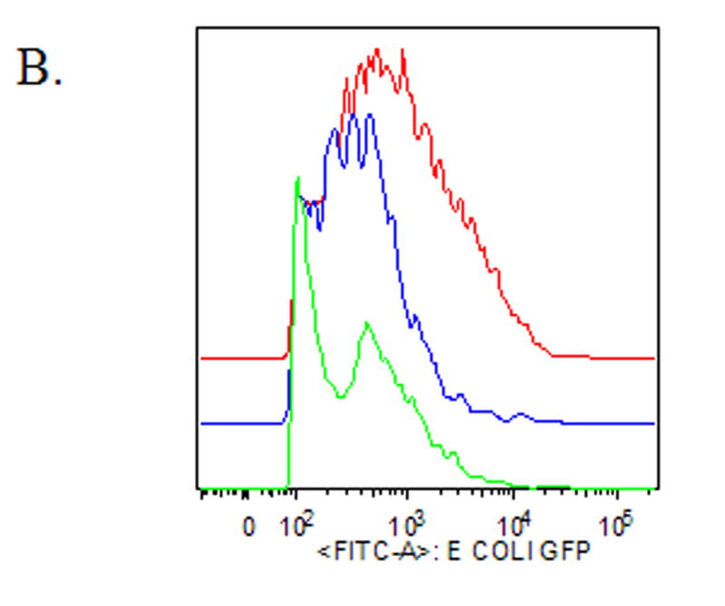
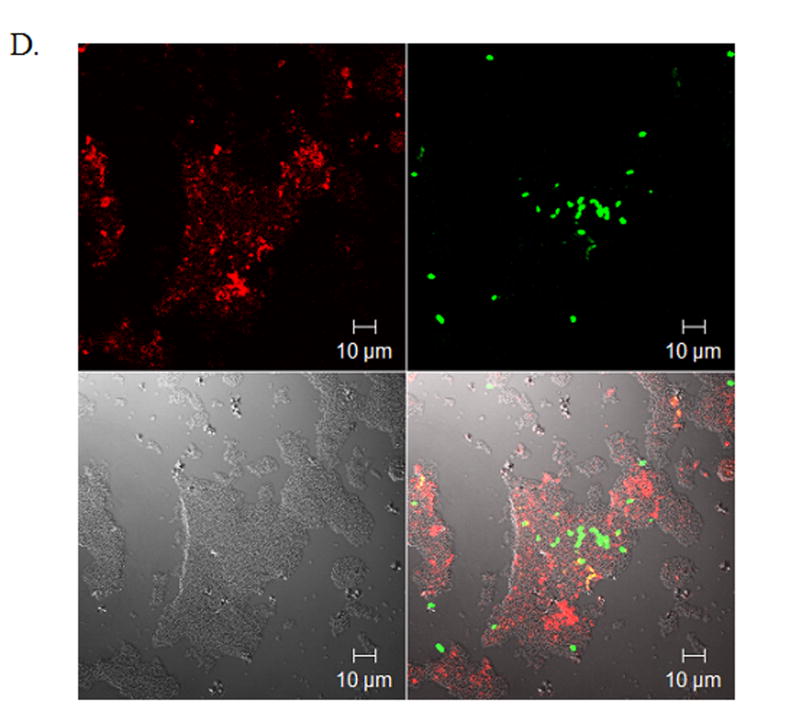
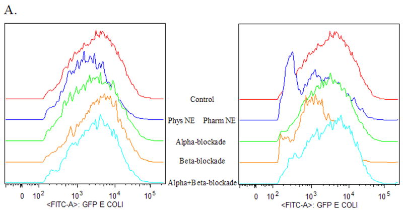
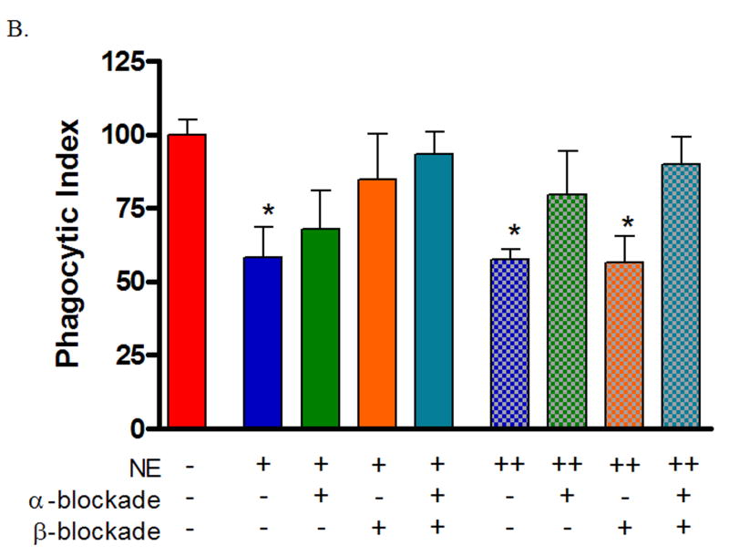
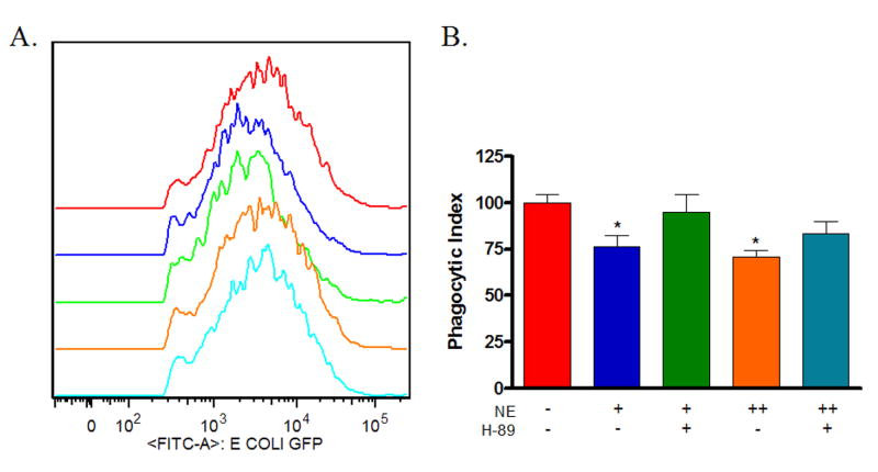
Methods: Murine wound macrophages were harvested at 5 days after injury and treated with physiologic and pharmacologic dose norepinephrine. Phagocytosis of green fluorescent protein-labeled Escherichia coli was assayed by flow cytometry. The signaling pathways mediating NE modulation of wound macrophage phagocytosis were interrogated by pharmacologic manipulation of alpha- and beta-adrenoreceptors (ARs), intracellular cyclic adenosine monophosphate (cAMP), and protein kinase A (PKA). Tissue specificity was determined by comparison of wound macrophages to splenic macrophages.

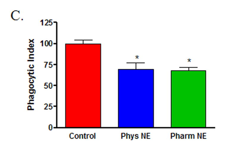
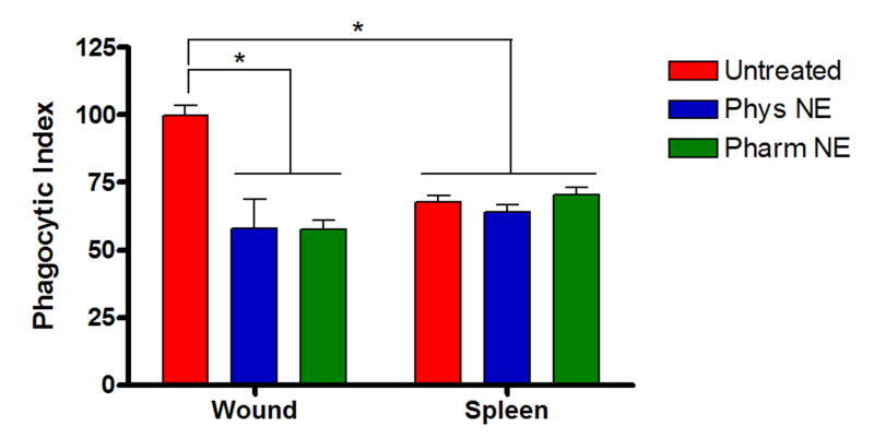
Results: Both physiologic and pharmacologic dose NE suppressed wound macrophage phagocytic efficiency. This effect was mediated by alpha- and beta-ARs in a dose-dependent fashion. Direct stimulation of cAMP-suppressed phagocytic efficiency and blockade of PKA signaling prevented NE-mediated suppression of phagocytic efficiency. Splenic macrophage phagocytic efficiency was less than that of wound macrophages and was not altered by NE.

Conclusions: NE has a profound immunosuppressive effect on wound macrophage function that is tissue specific and appears to be mediated through adrenergic receptors and their canonical downstream signaling pathway. Attenuation of post-injury immunosuppression represents another potential mechanism by which beta-AR blockade may reduce morbidity and mortality after severe injury.


Figures

Figure 1
Figure 1. Norepinephrine suppresses wound macrophage phagocytic efficiency
Macrophages isolated from 120-hour wounds were placed in culture for 18 hours with media alone (Control, red), physiologic norepinephrine (Phys NE, 10−9 M, blue), or pharmacologic norepinephrine (Pharm NE, 10−6 M, green) and phagocytosis of E.coli was assessed. (A) Norepinephrine-treatment did not alter the percentage of macrophages that engaged in phagocytosis (p=NS). (B) Representative phagocytosis histograms for control (red), physiologic NE-treated (blue) and pharmacologic NE-treated (green) wound macrophages. Both physiologic- (blue) and pharmacologic-dose norepinephrine treatment (green) resulted in a shift of the phagocytosis curve to the left, indicating fewer bacteria ingested per cell. The geometric mean of each curve was calculated to provide the phagocytic index (C) and was analyzed by two-way ANOVA followed by Bonferonni post-comparison testing. Both physiologic- and pharmacologic-dose norepinephrine suppressed the phagocytic efficiency of 120-hour wound macrophages (*p<0.001). (D) Representative confocal microscopy image demonstrating phagocytosis of GFP-E.coli (green, upper right) by a macrophage (phycoerythrin-F4/80, red, upper left). False-DIC (white, lower left) delineates the boundaries of the macrophage, demonstrating that the visualized bacteria are intracellular (merged image, lower right).
Figure 2
Figure 2. Norepinephrine-mediated suppression of wound macrophage phagocytosis is tissue-bed specific
Splenic macrophages isolated from animals 120-hours post-wounding were treated with physiologic norepinephrine (Phys NE, 10−9 M), or pharmacologic norepinephrine (Pharm NE, 10−6 M) for 18 hours and phagocytosis of E.coli was assessed in comparison to wound macrophages from the same animals. Splenic macrophage phagocytic function was less than that of wound macrophages at baseline(*p<0.001). Additionally, unlike wound macrophages (*p<0.001), norepinephrine treatment did not suppress splenic macrophage phagocytosis (p=NS).
Figure 3
Figure 3. Norepinephrine-mediated suppression of wound macrophage phagocytosis is adrenoreceptor dependent
Macrophages isolated from 120-hour wounds were pre-treated with either α- or β-adrenergic blockade for two hours prior to treatment with physiologic norepinephrine (NE +, 10−9 M) or pharmacologic norepinephrine (NE ++, 10−6 M) for 18 hours and assessment of phagocytosis of E.coli. Neither α- nor β-adrenergic blockade alone altered wound macrophage phagocytosis (p=NS, data not shown). α-adrenergic blockade partially prevented the suppression of wound macrophage phagocytosis by physiologic norepinephrine and completely blocked pharmacologic norepinephrine-mediated suppression (p=NS vs. Untreated Control). β-adrenergic blockade completely blocked physiologic norepinephrine-mediated suppression (p=NS) but did not prevent the suppression of wound macrophage phagocytosis by pharmacologic norepinephrine (*p<0.05). Combined α- and β-adrenergic blockade prevented both physiologic and pharmacologic norepinephrine-mediated suppression of wound macrophage phagocytosis (p=NS).
Figure 4
Figure 4. The adenylate cyclase activator forskolin suppresses wound macrophage phagocytosis
Macrophages isolated from 120-hour wounds were treated with physiologic norepinephrine, pharmacologic norepinephrine, or forskolin in the presence of phosphodiesterase inhibitor (IBMX) for 18 hours and phagocytosis of E.coli was assessed. Treatment with forskolin suppressed wound macrophage phagocytic efficiency(*p<0.05), although to a lesser degree than treatment with physiologic or pharmacologic doses of norepinephrine (#p<0.05).
Figure 5
Figure 5. Norepinephrine-mediated suppression of wound macrophage phagocytosis is mediated by protein kinase A (PKA)
Macrophages isolated from 120-hour wounds were treated with the PKA inhibitor H-89 for two hours prior to treatment with physiologic norepinephrine (NE +, 10−9 M) or pharmacologic norepinephrine (NE ++, 10−6 M) for 18 hours and assessment of phagocytosis of E.coli. PKA blockade alone had no effect on wound macrophage phagocytosis (p=NS, data not shown). PKA inhibition prevented physiologic norepinephrine-mediated suppression of wound macrophage phagocytosis and partially prevented pharmacologic norepinephrine-mediated suppression (p=NS vs. Untreated Control).
Figure 6
Figure 6. Putative model of norepinephrine-mediated alterations in wound macrophage phagocytosis
A putative model of norepinephrine-mediated modulation of wound macrophage phagocytic efficiency is proposed. At physiologic doses of norepinephrine, extracellular effects appear to be mediated primarily by β-adrenergic receptor binding (heavy line), with a smaller contribution from α-adrenergic receptor stimulation (light line). At the pharmacologic dose of norepinephrine, the reverse is true; the extracellular effects appear to be mediated primarily by α-adrenergic receptor binding (heavy line), with a smaller contribution from β-adrenergic receptor stimulation (light line). Regardless of dose, the intracellular alterations leading to decreased phagocytic efficiency involve increased levels of cAMP and its downstream effector, PKA.

Similar articles

Cited by

References

    1. Centers for Disease Control and Prevention: National Center for Injury Prevention and Control. Web-based Injury Statistics Query and Reporting System (WISQARS) www.cdc.gov/ncipc/wisqars. 2005. [cited 2006 May 4]; Available from:
    1. Baker CC, Oppenheimer L, Stephens B, Lewis FR, Trunkey DD. Epidemiology of trauma deaths. Am J Surg. 1980;140(1):144–50. - PubMed
    1. Deitch EA. Infection in the compromised host. Surg Clin North Am. 1988;68(1):181–97. - PubMed
    1. Elenkov IJ, Wilder RL, Chrousos GP, Vizi ES. The sympathetic nerve--an integrative interface between two supersystems: the brain and the immune system. Pharmacol Rev. 2000;52(4):595–638. - PubMed
    1. Woolf PD, McDonald JV, Feliciano DV, Kelly MM, Nichols D, Cox C. The catecholamine response to multisystem trauma. Arch Surg. 1992;127(8):899–903. - PubMed

Publication types

MeSH terms