

Polymicrobial sepsis enhances clearance of apoptotic immune cells by splenic macrophages
- PMID: 17689693
- PMCID: PMC2023968
- DOI: 10.1016/j.surg.2007.04.005
Polymicrobial sepsis enhances clearance of apoptotic immune cells by splenic macrophages
Abstract
Background: Macrophage phagocytosis of apoptotic cells induces an anti-inflammatory macrophage phenotype. Immune cell apoptosis is widespread in sepsis; however, it is unknown whether sepsis alters the capacity of macrophages to clear this expanded population. We hypothesize that sepsis will enhance splenic macrophage phagocytosis of apoptotic immune cells, potentially contributing to immunosuppression.
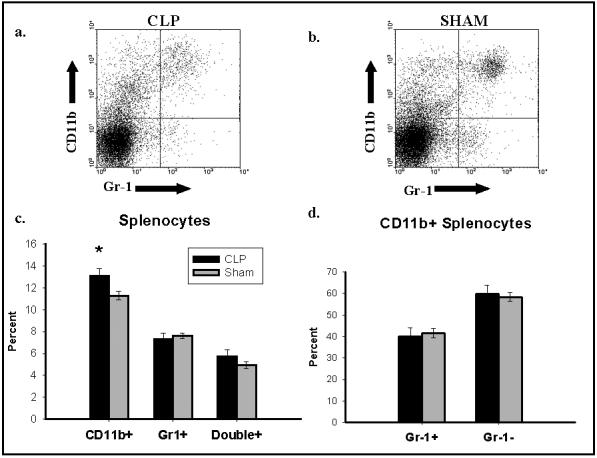
Methods: Sepsis was induced in C57BL/6J mice by cecal ligation and puncture (CLP). Apoptosis was induced in mouse thymocytes by dexamethasone incubation. At multiple time points after CLP/sham, splenic and peritoneal macrophages were isolated, plated on glass coverslips, co-incubated with apoptotic thymocytes, and fixed and the coverslips were then Giemsa stained. Splenic macrophages were also isolated 48 hours after CLP/sham, stained with the red fluorescent dye PKH26, and co-incubated with green fluorescent dye CFSE-stained apoptotic thymocytes and then coverslips were fixed and counterstained with DAPI. The macrophage phagocytic index (PI) was calculated for both staining methods.
Results: The PI of CLP splenic macrophages was significantly higher than sham by 24 hours, and this difference was sustained through 48 hours.
Conclusions: Studies suggest that apoptotic cell clearance leads to an anti-inflammatory macrophage condition, which together with our findings in septic macrophages, may point at a process that contributes to septic immune suppression.
Figures


References
-
- Angus DC, Linde-Zwirble WT, Lidicker J, Clermont G, Carcillo J, Pinsky MR. Epidemiology of severe sepsis in the United States: analysis of incidence, outcome, and associated costs of care. Crit Care Med. 2001;29:1303–1310. - PubMed
-
- Bone RC, Balk RA, Cerra FB, Dellinger RP, Fein AM, Knaus WA, Schein RM, Sibbald WJ. Definitions for sepsis and organ failure and guidelines for the use of innovative therapies in sepsis. Chest. 1992;101:1644–1655. - PubMed
-
- Hotchkiss RS, Karl IE. The pathophysiology and treatment of sepsis. N Engl J Med. 2003;348:138–150. - PubMed
-
- Wesche DE, Lomas-Neira JL, Perl M, Chung CS, Ayala A. Leukocyte apoptosis and its significance in sepsis and shock. J Leukocyte Biol. 2005;25:325–337. - PubMed
-
- Oberholzer C, Oberholzer A, Clare-Salzler M, Moldawer LL. Apoptosis in sepsis: a new target for therapeutic exploration. FASEB J. 2001;15:879–892. - PubMed
MeSH terms
Substances
Grants and funding
LinkOut - more resources
Full Text Sources
Medical
Research Materials
Miscellaneous
