. 2007 Aug;142(2):295-302.
doi: 10.1016/j.surg.2007.04.018.

Phosphatidylinositol 3-kinase pathway regulates hypoxia-inducible factor-1 to protect from intestinal injury during necrotizing enterocolitis

Affiliations

Phosphatidylinositol 3-kinase pathway regulates hypoxia-inducible factor-1 to protect from intestinal injury during necrotizing enterocolitis

Naira Baregamian et al. Surgery. 2007 Aug.

Abstract

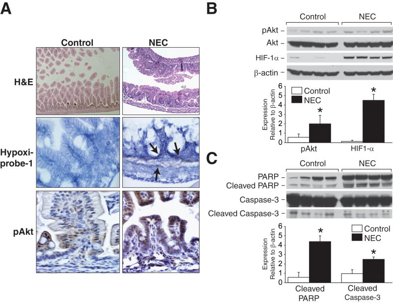
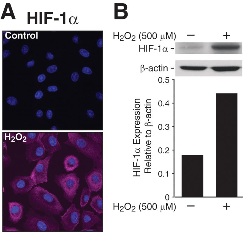
Background: Hypoxia is an important risk factor for development of necrotizing enterocolitis (NEC) in premature infants. Hypoxia-inducible factor (HIF)-1 is a transcription factor that plays a critical role in cellular responses to hypoxia and can be induced by phosphatidylinositol 3-kinase (PI3-K) pathway. Activation of the PI3-K and regulation of HIF-1 during NEC have not been elucidated.

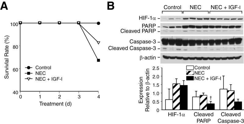
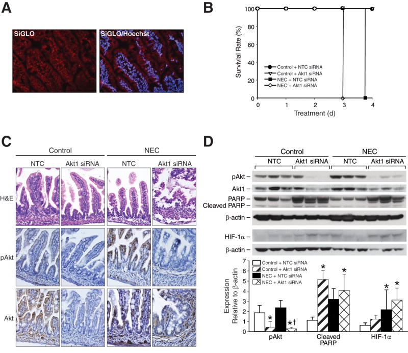
Methods: NEC was induced in 3-day-old neonatal mice using hypoxia and artificial formula feedings. Mice were divided into 3 treatment groups: (1) NEC alone, (2) NEC with insulin-like growth factor (IGF)-I, or (3) NEC with Akt1 siRNA treatment. Animals were sacrificed, and intestinal sections were harvested for protein analysis, H&E, and immunohistochemical staining.

Results: In vivo model of NEC produced intestinal injury associated with increased protein expression of HIF-1alpha, pAkt, PARP, and caspase-3 cleavage. Pretreatment with IGF-1 attenuated an HIF-1alpha response. In contrast, targeted inhibition of Akt1 completely abolished NEC-induced expression of pAkt and upregulated HIF-1alpha activation.

Conclusions: NEC activates important protective cellular responses to hypoxic injury such as HIF-1alpha and PI3-K/Akt in neonatal gut. Hypoxia-mediated activation of pro-survival signaling during NEC may be modulated with growth factors, which thus suggests a potential therapeutic option in the treatment of neonates with NEC.


Figures

Fig 1
Fig 1. In vivo NEC activates PI3-K/Akt pathway and increases HIF-1α expression
(A) Blunting of villous tips, submucosal separation/edema are present in NEC sections (first row). Hypoxia is markedly expressed in intestinal lamina propria and submucosa during NEC (second row; arrows). Increased intestinal pAkt expression is noted during in vivo NEC (third row). (B) Extracted total proteins from ileal segments were analyzed by Western blotting for the expression of HIF-1α, pAkt and total Akt. NEC induced significant increase in pAkt and HIF-1α expression (* p < 0.05 vs. control). Equal β-actin levels indicate even loading. (C) NEC intestinal tissues were harvested and analyzed for apoptotic protein markers, PARP and caspase-3. NEC resulted in significantly increased cleavage of PARP and caspase-3 (* p < 0.05 vs. control).
Fig 2
Fig 2. H2O2 treatment induces HIF-1α expression in RIE-1 cells
(A) RIE-1 cells (1×10⁴/well) were treated with H2O2 (500 μM) for 1h, and then incubated with HIF-1α antibody, AlexaFluor® 647-labeled goat anti-rabbit IgG and Höechst 33342 nuclear stain. They were visualized using 1.0 Zeiss LSM 510 UV Meta laser scanning confocal microscopy. H2O2 treatment resulted in significant increase in intracellular HIF-1α activation and accumulation by nearly 10-fold in RIE-1 cells. (Zeiss LSM Image software). (B) RIE-1 cells were treated with H2O2 for 1h, the protein was extracted for Western blot analysis. H2O2 treatment also increased protein expression of HIF-1α. Equal β-actin levels indicate even loading.
Fig 3
Fig 3. IGF-1 treatment improves mice survival during NEC and attenuates apoptosis, HIF-1α expression during NEC
(A) Neonatal mice were randomized into control (n=5), NEC alone (n=6) and IGF-1 + NEC (n=6) groups, and then sacrificed after 96 h. Neonatal mice in a NEC group with single IGF-1 injection (10 μg/g; i.p.) showed higher survival rate than pups in NEC alone group (83.3% vs. 66.6%). (B) Ileal segments were harvested and analyzed by Western blotting for the expression of intestinal HIF-1α, caspase-3 and PARP († p < 0.05, NEC alone vs. NEC + IGF-1). IGF-1 pretreatment attenuated NEC-induced cleavage of PARP and caspase-3, and decreased HIF-1α protein levels. Equal β-actin levels indicate even loading.
Fig 4
Fig 4. Akt1 siRNA induces HIF-1α upregulation and decreases survival during NEC
(A) Intestinal cryosections from healthy 3-day old mice (n=3) injected with siGLO Lamin A/C siRNA i.p. were stained with Höechst 33342 nuclear stain. Intestinal sections showed excellent siGLO Lamin A/C siRNA transfection and localization in neonatal mouse intestine (Nikon fluorescent microscopy). (B) Neonatal mice were randomized into control and NEC groups to receive either NTC or Akt1 siRNA i.p injections for 96 h. Five mice were used for each treatment group. Neonatal mice in a NEC Akt1 siRNA group had significantly higher mortality rate than pups in NEC NTC siRNA. (C) Intestinal sections stained with H&E showed mucosal and submucosal injury in NEC NTC and NEC Akt1 siRNA groups (first row). Intestinal pAkt and Akt levels were attenuated in Akt1 siRNA-treated groups during NEC (second and third rows). (D) Ileal segments were harvested and analyzed by Western blotting for the expression of intestinal HIF-1α, pAkt, Akt1 and PARP. Targeted Akt1 inhibition enhaced NEC-induced HIF-1α protein levels, enhanced PARP cleavage, and effectively silenced activation of Akt1 in control and NEC groups (* p < 0.05 vs. control; † p < 0.05, NEC Akt1 siRNA vs. NEC NTC siRNA). Equal β-actin levels indicate even loading.

References

    1. Sibbons P, Spitz L, van Velzen D, Bullock GR. Relationship of birth weight to the pathogenesis of necrotizing enterocolitis in the neonatal piglet. Pediatr Pathol. 1988;8:151–62. - PubMed
    1. Schumacker PT. Hypoxia-inducible factor-1 (HIF-1) Crit Care Med. 2005;33:S423–5. - PubMed
    1. Zundel W, Schindler C, Haas-Kogan D, Koong A, Kaper F, Chen E, et al. Loss of PTEN facilitates HIF-1-mediated gene expression. Genes Dev. 2000;14:391–6. - PMC - PubMed
    1. Zhong H, Chiles K, Feldser D, Laughner E, Hanrahan C, Georgescu MM, et al. Modulation of hypoxia-inducible factor 1alpha expression by the epidermal growth factor/phosphatidylinositol 3-kinase/PTEN/AKT/FRAP pathway in human prostate cancer cells: implications for tumor angiogenesis and therapeutics. Cancer Res. 2000;60:1541–5. - PubMed
    1. Chandel NS, Maltepe E, Goldwasser E, Mathieu CE, Simon MC, Schumacker PT. Mitochondrial reactive oxygen species trigger hypoxia-induced transcription. Proc Natl Acad Sci U S A. 1998;95:11715–20. - PMC - PubMed

Publication types

MeSH terms

Substances