Clinical Trial
. 2007 Nov;48(2):87-101.
doi: 10.1016/j.jdermsci.2007.06.006. Epub 2007 Aug 8.

Targeting TNFalpha rapidly reduces density of dendritic cells and macrophages in psoriatic plaques with restoration of epidermal keratinocyte differentiation

Affiliations
Clinical Trial

Targeting TNFalpha rapidly reduces density of dendritic cells and macrophages in psoriatic plaques with restoration of epidermal keratinocyte differentiation

Deborah J Marble et al. J Dermatol Sci. 2007 Nov.

Abstract

Background: The cytokine network theory for psoriasis postulates a key role for TNFalpha in mediating inflammation and altered epidermal differentiation.

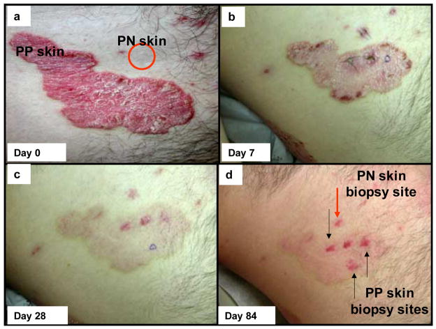
Objective: This study defines responses following administration of adalimumab, a TNFalpha inhibitor, in pre-psoriatic skin (PN) and lesional psoriatic plaques (PP) skin.

Methods: PN and PP skin before and after treatment were biopsied at days 2, 7, 28 and 84 (n=6 different patients). Cryosections were immunohistochemically stained to detect TNFalpha and other relevant markers in epidermal and dermal compartments. Detection of apoptosis utilized antibody specific for activated caspase 3. Semiquantitative assessments and statistical analysis was performed for each staining profile.

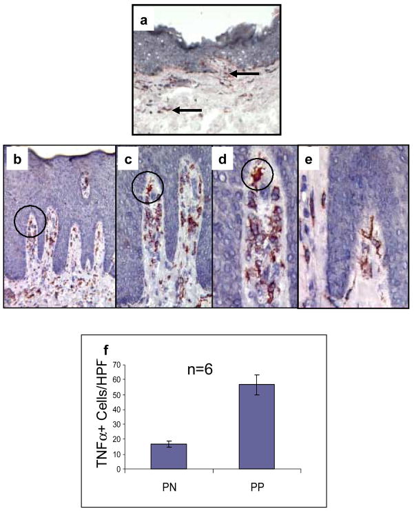
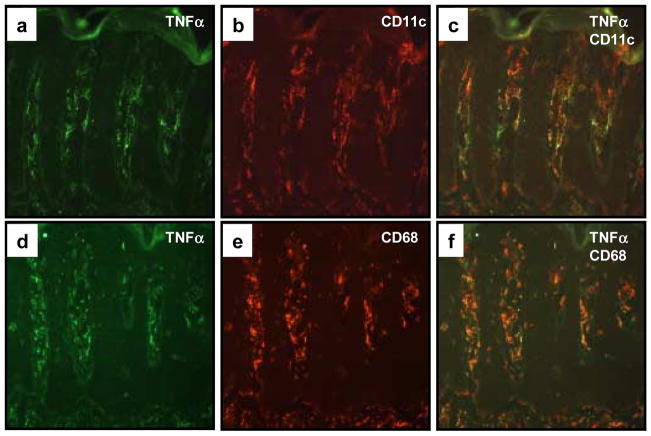
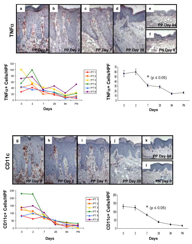
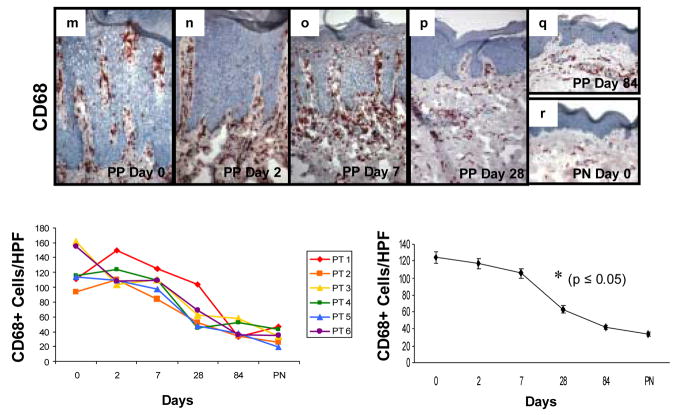
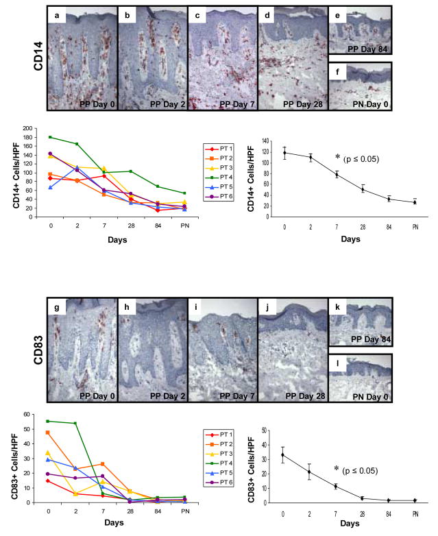
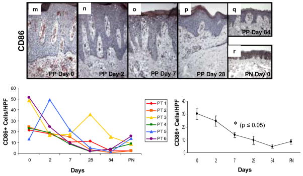
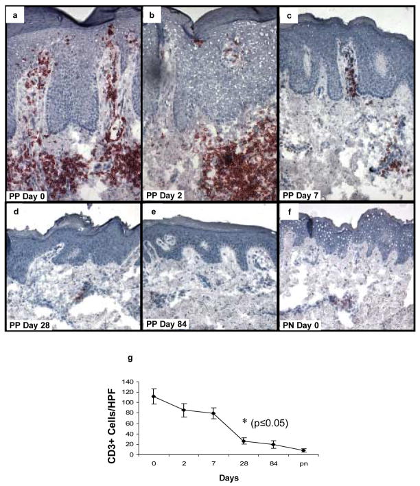
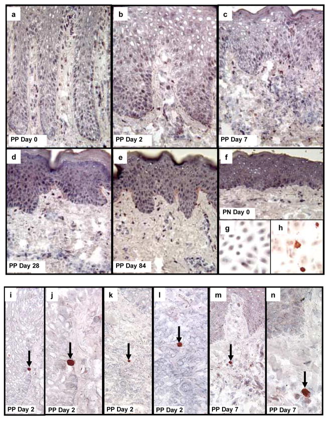
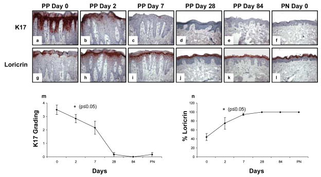
Results: TNFalpha+ cells were increased in PP skin. PP skin was also characterized by a four-fold increase in number of CD68+ macrophages as well as eight-fold increase in CD11c+ dermal dendritic cells (DCs) compared to PN skin. By two-color immunofluorescence staining, both CD68+ cells as well as CD11c+ cells expressed TNFalpha. Following initiation of adalimumab therapy, CD11c+ cells, significantly decreased in PP skin at days 7, 28, and 84, while CD68+ and CD14+ cells decreased at days 28 and 84. Other markers for DCs (CD83, CD86) showed decreases at days 7, 28, and 84. Reduction in DCs, macrophages or T cells was not accompanied by increased activated caspase 3-positive cells. When a keratinocyte terminal differentiation marker was examined, adalimumab triggered rapid restoration of loricrin expression (beginning on day 2), with loss of aberrant differentiation marker, keratin 17 (K17).

Conclusion: Adalimumab impacts dermal-based immunocytes, and the epidermal compartment also responds by restoration of normal differentiation without detectable apoptosis.


Figures

Figure 1
Figure 1. Clinical perspective of skin lesions on a psoriatic patient before and after adalimumab therapy
Compared to adjacent symptomless (PN) skin, designated by red circle, the baseline appearance for untreated psoriatic plaques (PP skin) revealed well demarcated, erythematous, and scaly lesions (a). On day 7 after delivery of a single loading dose of adalimumab (80 mg, subcutaneous) the lesional skin is characterized by reduced thickness, erythema, and scaling (b; the sutures represent biopsy sites for PN and PP skin). On day 28 after continuation of adalimumab therapy, the target plaque is almost completely resolved with only slight residual erythema; a clinical trend continued on day 84 of therapy (c, d). Note the resolution of the inflammation and tissue remodeling of PP skin by adalimumab was not accompanied by any scar formation.
Figure 2
Figure 2. TNFα expression in PN and PP skin
Cryopreserved sections of PN skin (panel a) and PP skin (panels be) were immunostained to detect TNFα. Note the markedly increased number of TNFα + cells in PP skin (b) compared to PN skin (a). The majority of TNFα + cells in PP skin are located in the papillary dermis (be). Note that progressively increased magnification is provided in panels bd using a black circle for orientation. Many of the TNFα + cells had a dendritic morphology (e), whereas others had a plump macrophage-like appearance (d). No epidermal keratinocyte immunoreactivity for TNFα was observed. Quantitation of TNFα immunoreactive cells involving six different subjects (n=6) for PN skin (17±2 cells/HPF) and PP skin (57±7 cells/HPF) revealed significant differences between PN and PP skin (p<0.05) as depicted in (f). These results indicate PP skin is characterized by an increased density of TNFα + mononuclear cells that infiltrate the papillary dermis and focally extend into the epidermal compartment.
Figure 3
Figure 3. Co-localization of TNFα expressing cells within CD11c+ dendritic cells and CD68+ macrophages
Cryopreserved sections of PP skin were subjected to two-color immunofluorescence staining in which TNFα expressing cells were labeled green (FITC; a, d), and the CD11c+ (b) or CD68+ (e) mononuclear cells labeled red (rhodamine). Note the similar distribution patterns and morphological profiles, as well as dual fluorescence imaging that highlight the co-expression of TNFα in both CD11c+ (c) as well as CD68+ (f) cell subsets.
Figure 4
Figure 4. Expression and kinetic profile highlighting temporal reductions in density of TNFα +, CD11c+, and CD68+ cells in psoriatic patients before and after treatment with adalimumab
Untreated PP skin (PP day 0) is characterized by an increased number of mononuclear cells expressing TNFα compared to PN skin (a and f), as well as increased number of CD11c+ cells (g and l), and CD68+ cells (m and r). Beneath each representative staining profile of untreated and treated PP skin and PN skin are panels depicting individual data points for each of the six treated subjects (left side), as well as a summary panel depicting mean +/− SEM for the group (n=6). For each treated subject, a color and symbol are provided to delineate a specific patient in figures 4 and 5. Note the consistent and rapid reduction in TNFα expressing cells (panels b–e, s) which first became statistically significant at day 7 (asterisk; p<0.05), as does the reduction in density of CD11c+ cells (panels h–k, t); whereas the reduction in CD68+ cells does not achieve significance until day 28 (panels n–q; u). Note the parallel reduction in epidermal thickness that accompanies the diminution in the density of mononuclear cells at the indicated time points, consistent with our previous report [24].
Figure 5
Figure 5. Expression and kinetic profile highlighting temporal reductions in density of CD14+, CD83+, and CD86+ cells in psoriatic patients before and after treatment with adalimumab
Untreated PP skin (PP day 0) is characterized by an increased number of mononuclear cells expressing CD14+ compared to PN skin (a and f), as well as increased number of DCs expressing maturation markers CD83 (g and l) and CD86 (m and r). Beneath each representative staining profile of untreated and treated PP skin and PN skin are panels depicting individual data points for each of the six treated subjects (left side), as well as a summary panel depicting mean +/− SEM for the group (n=6). The reduction in these markers initially became statistically significant at day 7 (asterisk; p<0.05 comparing untreated to treated samples).
Figure 6
Figure 6. Expression and kinetic profile highlighting temporal reductions in density of CD3+ T cells in psoriatic patients before and after treatment with adalimumab
Untreated PP skin is characterized by influx of CD3+ T cells, located both in the upper dermis with focal extension into the epidermis. Following treatment with adalimumab, the number of T cells begins to decline on days 2 and 7 (b, c), with a more pronounced reduction on days 28 and 84 (d, e) where they resemble the density in PN skin (f). The earliest time point with statistically significant reduction in CD3+ cell counts compared to untreated samples is day 28 (g; n=6; asterisk, p<0.05).
Figure 7
Figure 7. Detection of activated caspase 3 as a marker for cells undergoing apoptosis in psoriatic patients before and after treatment with adalimumab
Untreated PP and PN skin are characterized by the absence of cells in either the epidermis or dermis undergoing apoptosis because no activated caspase 3 positive cells are present in the tissue samples (a, f). Moreover, following treatment with adalimumab, the reduction in epidermal thickness as well as the decreased density of immunocytes (dendritic cells, macrophages, T cells) was also not accompanied by and prominent increase in detectible cells expressing activated caspase 3 (be). As a positive control, cultured human keratinocytes were immunostained before (g), and after, (h) UV-light to confirm detection of activated caspase 3. Note the strong cytoplasmic immunostaining for activated caspase 3 in keratinocytes undergoing apoptosis following UV-light (h). In skin tissue sections, occasional, rare scattered cells expressing bright red anti-activated caspase 3 reaction product was detected in epidermal keratinocytes (i, j; arrows depict basal layer keratinocyte undergoing apoptosis in PP skin 2 days after adalimumab treatment. A rare dermal lymphocyte expressing activated caspase 3 was also identified at day 2 in treated PP skin (k, l; arrows depict apoptotic lymphocyte). A rare macrophage-like cell expressing activated caspase 3 is shown in (m, n) on day 7 in treated PP skin.
Figure 8
Figure 8. Expression and kinetic profile highlighting epidermal response in psoriatic patients before and after treatment with adalimumab focusing on aberrant keratin 17 and loricrin expression
Untreated PP skin is characterized by strong and diffuse epidermal keratinocyte expression of the aberrant differentiation marker, keratin 17 (K17; a), accompanied by reduced expression of the terminal differentiation marker, loricrin (g). As early as 2 days following a single injection of adalimumab (n=6), there is significant reduction in K17 expression (b) with restoration of strong diffuse loricrin expression (h). By day 7, there is almost complete loss of K17 expression (c) with continued normalization of loricrin expression and orthokeratosis (h). Biopsies on day 28 and 84 revealed near complete healing of PP skin resembling PN skin as depicted (d–f, j–l).

Similar articles

Cited by

References

    1. Krueger JG. The immunologic basis for the treatment of psoriasis with new biologic agents. J Am Acad Dermatol. 2002;46:1–23. - PubMed
    1. McKay IA, Leigh IM. Altered keratinocyte growth and differentiation in psoriasis. Clin Dermatol. 1995;13:105–114. - PubMed
    1. Nickoloff BJ, Nestle FO. Recent insights into the immunopathogenesis of psoriasis provide new therapeutic opportunities. J Clin Invest. 2004;113:1664–1675. - PMC - PubMed
    1. Austin LM, Ozawa M, Kikuchi T, Walters IB, Krueger JG. The majority of epidermal T cells in Psoriasis vulgaris lesions can produce type 1 cytokines, interferon-gamma, interleukin-2, and tumor necrosis factor-alpha, defining TC1 (cytotoxic T lymphocyte) and TH1 effector populations: a type 1 differentiation bias is also measured in circulating blood T cells in psoriatic patients. J Invest Dermatol. 1999;113:752–759. - PubMed
    1. Lew W, Bowcock AM, Krueger JG. Psoriasis vulgaris: cutaneous lymphoid tissue supports T-cell activation and “Type 1” inflammatory gene expression. Trends Immunol. 2004;25:295–305. - PubMed

Publication types

MeSH terms