. 2007;35(16):5312-22.
doi: 10.1093/nar/gkm493. Epub 2007 Aug 8.

Phospho-epitope binding by the BRCT domains of hPTIP controls multiple aspects of the cellular response to DNA damage

Affiliations

Phospho-epitope binding by the BRCT domains of hPTIP controls multiple aspects of the cellular response to DNA damage

Ivan M Munoz et al. Nucleic Acids Res. 2007.

Erratum in

Expression of concern in

Abstract

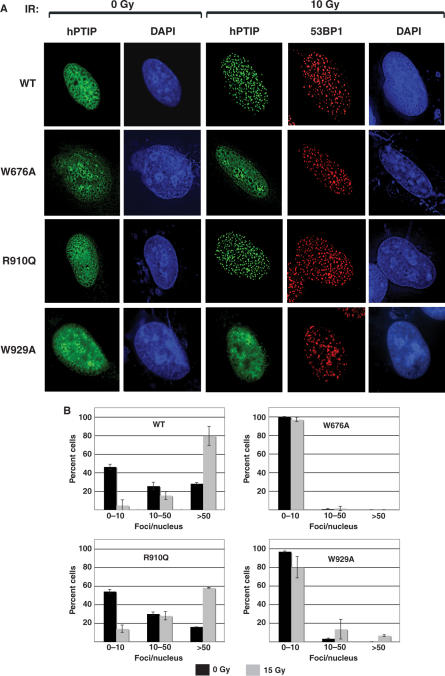
Human (h)PTIP plays important but poorly understood roles in cellular responses to DNA damage. hPTIP interacts with 53BP1 tumour suppressor but only when 53BP1 is phosphorylated by ATM after DNA damage although the mechanism(s) and significance of the interaction of these two proteins are unclear. Here, we pinpoint a single ATM-phosphorylated residue in 53BP1--Ser25--that is required for binding of 53BP1 to hPTIP. Binding of phospho-Ser25 to hPTIP in vitro and in vivo requires two closely apposed pairs of BRCT domains at the C-terminus of hPTIP and neither pair alone can bind to phospho-Ser25, even though one of these BRCT pairs in isolation can bind to other ATM-phosphorylated epitopes. Mutations in 53BP1 and in hPTIP that prevent the interaction of the two proteins, render cells hypersensitive to DNA damage and weaken ATM signalling. The C-terminal BRCT domains of hPTIP are also required for stable retention of hPTIP at sites of DNA damage but this appears to be independent of binding to 53BP1. Thus, the BRCT domains of hPTIP play important roles in the cellular response to DNA damage.


Figures

Figure 1.
Figure 1.
Identification of Ser25 of 53BP1 as the ATM-phosphorylated residue required for interaction with hPTIP after IR. (A) Schematic diagram of full-length 53BP1. Asterisks represent S/T-Q motifs. The TUDOR domains and BRCT domains of 53BP1 are shown in green and blue, respectively. In MUT-C10, the serine or threonine residue in each of the 10 most C-terminal S/T-Q motifs were mutated to alanine. In MUT-N17, the 17 S/T-Q motifs immediately upstream of those in MUT-C10 were mutated. In MUT-N20, a further three S/T-Q motifs were mutated in addition to those in MUT-N17. (BD) HEK293 cells were co-transfected with FLAG-hPTIP and full-length HA-53BP1 bearing the indicated mutations. Cells were exposed to IR (0 Gy or 20 Gy) and after cell lysis anti-FLAG immunoprecipitates were subjected to SDS–PAGE and western blotting with the indicated antibodies. The lowest panel in each case shows HA-53BP1 levels in cell extracts. (E) HEK293 cells were pre-incubated with DMSO vehicle or with 10 μM KU55933 or 10 μM NU7441 for 1 h before exposure to IR (10 Gy) and then allowed to recover for the times indicated. Cells were lysed in LDS sample and extracts subjected to SDS–PAGE followed by western blot analysis with the indicated antibody. (F) HEK293 cells grown on 13-mm-diameter glass coverslips were transfected with full-length HA-53BP1: wild-type (WT) or the Ser25Ala mutant and then treated with 0 or 10 Gy IR and left to recover at 37°C for 45 min before fixation and permeabilization as described previously (3). Cells were washed and blocked before incubation with anti-HA (to detect transfected 53BP1).
Figure 2.
Figure 2.
Both C-terminal BRCT domains of hPTIP mediate binding to phospho-Ser25 of 53BP1. (A) Schematic diagram of hPTIP. BRCT domains are shown as blue boxes. (B) Extracts from untransfected HEK 293 cells (top panel; 4 mg of extract protein), or cells transfected with full-length FLAG-hPTIP (middle panel; 100 μg of extract protein) or with FLAG-hPTIP (590–1069; 100 μg of extract protein) corresponding to the two C-terminal BRCT domains of hPTIP (‘Pair C1 + Pair C2’; bottom panel) were incubated with Dynabeads conjugated to the indicated peptides, corresponding to the sequence around Ser25 and Ser29 of 53BP1. The peptides used were: Empty, no peptide; Non(P), biotin-DTPCLIIEDSQPEQVLEDD; Ser25(P), biotin-DTPCLIIEDpSQPESQVLEDD; Ser29(P), biotin-DTPCLIIEDSQPEpSQVLEDD; Ser25(P)/Ser29(P), biotin-DTPCLIIEDpSQPEpSQVLEDD; Rad53(P), biotin-MENIpTQPpTQQSTQAT. (pS, phosphoserine, pT, phosphothreonine). After extensive washing, beads were subjected to SDS–PAGE and western blot analysis with the indicated antibodies. (C) Bacterially expressed GST-hPTIP (590–1069) (BRCT pairs C1 + C2), GST-hPTIP (590–800) (BRCT pair C1) and GST-hPTIP (862–1069) (BRCT pair C2) were incubated with Dynabeads conjugated to the indicated peptides (Figure 2B). After extensive washing, beads were subjected to SDS–PAGE and gels were stained with Coomassie brilliant blue. (D) Same as C. except that Non(P) refers to the peptide biotin-GAAYDI-SQ-VFPFAKKK while S-Q-V-F (P) refers to the peptide biotin-GAAYDI-pSQ-VFPFAKKK (4). Rad53(P) is described in (B).
Figure 3.
Figure 3.
BiaCore analysis of binding of hPTIP BRCT domain pairs C1 + C2 to 53BP1 phospho-Ser25. (A) The binding of bacterially expressed hPTIP (590–1069) (BRCT pairs C1 + C2) to biotinylated 53BP1 peptides (Non(P), biotin-DTPCLIIEDSQPEQVLEDD; Ser25(P), biotin-DTPCLIIEDpSQPESQVLEDD) was analysed using BiaCore as described in the Materials and Methods section. The analysis was performed over a range of protein concentrations (7.8–500 nM) and the response level at steady state was plotted against protein concentration. The Kd values were calculated by fitting the data to the equation y = Bmax·x/(Kd + x) using GraphPad 4 software, which describes the binding of a ligand to a receptor that follows the law of mass action. Bmax is the maximal binding and Kd is the concentration of ligand required to reach half-maximal binding, whereas x and y correspond to the protein concentration and the response units, respectively. (B) Same as A., except that binding of hPTIP BRCT domain pair C1, C2 and pair C1 + pair C2 to the 53BP1 phospho-Ser25 peptide was compared.
Figure 4.
Figure 4.
Effect of mutation of conserved residues in the hPTIP BRCT domains on binding to phospho-53BP1 (A) The binding of bacterially expressed hPTIP-BRCT pair C1 + C2 proteins: wild type, W676A, R910Q and W929A to the 53BP1 phospho-Ser25 peptide (biotin-DTPCLIIEDpSQPESQVLEDD) was analysed using BiaCore as described in the Materials and Methods section and in the legend to Figure 3A. The inset shows a Coomassie-stained gel of the GST fusion proteins used in this analysis. (B) HEK293 cells were co-transfected with full-length HA-53BP1 and FLAG-hPTIP bearing the indicated mutations. Cells were exposed to IR (0 Gy or 20 Gy) and after cell lysis anti-FLAG immunoprecipitates were subjected to SDS–PAGE and western blotting with the indicated antibodies. The lowest panel in each case shows HA-53BP1 levels in cell extracts. (C) Bacterially expressed versions of GST-hPTIP-BRCT pair C1 + C2: wild-type (WT) and Trp676Ala (W676A), GST-hPTIP-BRCT pair C1 and GST-hPTIP-BRCT pair C2, or GST alone were immobilized on glutathione-sepharose and incubated with extracts of HEK 293 cells that had been exposed, or not, to 10 Gy IR. After extensive washing, beads were subjected to SDS–PAGE and western blot analysis with antibodies against 53BP1. Low and high exposures of the blot are shown in the top and middle panels, respectively and the bottom panel shows a Coomassie-stained gel of the GST-fusions used in this experiment. NS, non-specific.
Figure 5.
Figure 5.
Interaction of 53BP1 with hPTIP is not required for stable retention of hPTIP at sites of DNA damage. (A) U2OS cells grown on 13-mm-diameter glass coverslips were transfected with full-length FLAG-hPTIP: wild-type (WT) or the W676A, R910Q or W929A mutants and then treated with 0 or 15 Gy IR and left to recover at 37°C for 45 min before fixation and permeabilization as described previously (3). Cells were washed and blocked before incubation with anti-FLAG (to detect transfected hPTIP) or with anti-53BP1 (1 µg/ml) for 60 min at room temperature. After washing in PBS-T, coverslips were incubated with secondary antibodies (2 µg/ml) conjugated to Alexa488 or Alexa 594 (Jackson Laboratories) for 60 min. After thorough washing in PBS-T, coverslips were mounted on glass slides and images were acquired using an Olympus IX70 microscope. (B) The number of PTIP foci formed before and after exposure of cells to IR, in (A), was quantitated by counting the number of foci observed with anti-FLAG antibody in ∼200 transfected cells. The mean number of foci ± SD are presented.
Figure 6.
Figure 6.
The interaction of 53BP1 with hPTIP is required for an intact cellular response to DNA damage. (A) HEK293 cells were co-transfected with siRNA to silence 53BP1 or a scrambled RNA duplex (SCR), and with various plasmids: pCMV5 (−), pCMV5-HA-53BP1 carrying a silent mutation (see Materials and Methods section) that renders HA-53BP1 refractory to silencing by siRNA (WT), or pCMV5-HA-53BP1 with the same mutation but with Ser25 mutated to alanine (S25A). Cells were exposed to IR (3 Gy) and allowed to recover for the indicated times before lysis. Extracts were subjected to SDS–PAGE and western blotting with the indicated antibodies. (B) Same as (A) except that cells were seeded at different dilutions, in triplicate in 10-cm culture dishes and irradiated with 0 or 3 Gy IR. Before plating, the viability of the cells was assessed during counting by a dye exclusion test with trypan blue. After 14 days, colonies were fixed with methanol, stained with crystal violet and were counted if they consisted of more than 50 cells. The fraction of cells surviving irradiation was normalized to the surviving fraction of the corresponding control. (C) Same as (A) except that cells were co-transfected with siRNA to silence hPTIP (3) or a scrambled RNA duplex (SCR), and with various plasmids: pCMV5 (−), pCMV5-FLAG-hPTIP carrying a silent mutation that renders hPTIP refractory to silencing by siRNA (WT), or pCMV5-FLAG-hPTIP with Arg910 mutated to glutamine (R910Q).

References

    1. Lechner MS, Levitan I, Dressler GR. PTIP, a novel BRCT domain-containing protein interacts with Pax2 and is associated with active chromatin. Nucleic Acids Res. 2000;28:2741–2751. - PMC - PubMed
    1. Cho EA, Prindle MJ, Dressler GR. BRCT domain-containing protein PTIP is essential for progression through mitosis. Mol. Cell. Biol. 2003;23:1666–1673. - PMC - PubMed
    1. Jowsey PA, Doherty AJ, Rouse J. Human PTIP facilitates ATM-mediated activation of p53 and promotes cellular resistance to ionizing radiation. J. Biol. Chem. 2004;279:55562–55569. - PubMed
    1. Manke IA, Lowery DM, Nguyen A, Yaffe MB. BRCT repeats as phosphopeptide-binding modules involved in protein targeting. Science. 2003;302:636–639. - PubMed
    1. Durocher D, Jackson SP. DNA-PK, ATM and ATR as sensors of DNA damage: variations on a theme? Curr. Opin. Cell. Biol. 2001;13:225–231. - PubMed

Publication types

MeSH terms