. 2007 Aug 21;104(34):13690-5.
doi: 10.1073/pnas.0705053104. Epub 2007 Aug 9.

Conserved P-TEFb-interacting domain of BRD4 inhibits HIV transcription

Affiliations

Conserved P-TEFb-interacting domain of BRD4 inhibits HIV transcription

Dwayne A Bisgrove et al. Proc Natl Acad Sci U S A. .

Abstract

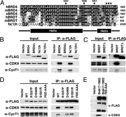
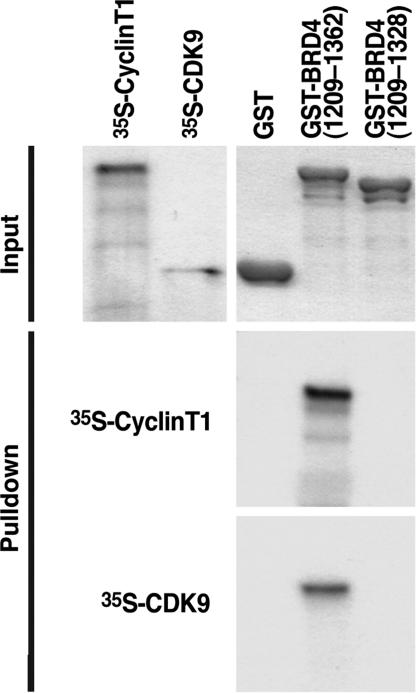
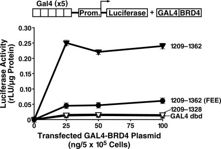
We have identified a conserved region in the C-terminal domain of bromodomain-containing protein 4 (BRD4) that mediates its specific interaction with positive transcription elongation factor b (P-TEFb). This domain is highly conserved in testis-specific bromodomain protein (BRDT) and Drosophila fs(1)h. Both BRDT and fs(1)h specifically interact with P-TEFb in mammalian cells, and this interaction depends on their C-terminal domains. Overexpression of the BRD4 P-TEFb-interacting domain disrupts the interaction between the HIV transactivator Tat and P-TEFb and suppresses the ability of Tat to transactivate the HIV promoter. Incubation of cells with a synthetic peptide containing the C-terminal domain of BRD4 interferes with transactivation of the HIV promoter by the Tat protein.


Conflict of interest statement

The authors declare no conflict of interest.

Figures

Fig. 1.
Fig. 1.
BRD4 interacts with P-TEFb by means of a short CTD. (A) FLAG-tagged deletion mutants of BRD4 were overexpressed in 293T cells. Nuclear extracts (NE) and anti-FLAG immunoprecipitates (IP) were immunoblotted (IB) for CDK9 and cyclin T1. The small arrows point to the full-length proteins, and the smaller bands likely reflect degradation products. (B) FLAG-tagged BRD4 constructs encoding residues 1209–1362, 1209–1362Δ1279–1304, 1209–1362Δ1306–1330, 1209–1362Δ1329–1362, 1209–1362Δ1329–1345, and 1209–1362Δ1345–1362 were overexpressed in 293T cells. Anti-FLAG immunoprecipitates were immunoblotted with anti-CDK9 or anti-cyclin T1.
Fig. 2.
Fig. 2.
A C-terminal peptide conserved among BRD4, BRDT, and fs(1)h mediates P-TEFb interaction. (A) Alignment of the C termini of human, murine, and Xenopus BRD4; human and murine BRDT; and Drosophila fs(1)h. Identical residues are indicated in black, and conserved residues are in gray. The locations of α-helices predicted by computer analysis are indicated below. (B) Vectors encoding the C termini of human BRD4(1209–1362), BRD4(Δ1209–1344), fs(1)h(1885–2038), or fs(1)hΔ(1885–2020) were transfected into 293T cells. Anti-FLAG immunoprecipitates (IP) were immunoblotted with anti-FLAG, anti-cyclin T1 (anti-CycT1), and anti-CDK9. (C) Expression constructs encoding the C terminus of BRDT (residues 821–956) or a C-terminal deletion mutant [BRDTΔ(821–938)] were transfected into 293T cells. Whole-cell lysates were prepared, and BRDT was immunoprecipitated with anti-FLAG beads. (D) C-terminal residues conserved among BRD4, BRDT, and fs(1)h were mutated in FLAG-BRD4(1209–1362) (indicated by an asterisk in A). Plasmids encoding BRD4 point mutants 1209–1362R1337P, D1346N, S1351R, and FEE1357AAA were transfected into 293T cells. Anti-FLAG immunoprecipitates were immunoblotted for FLAG, CycT1, and CDK9. (D) Plasmids encoding FLAG tagged wild-type (WT) BRD4 (1–1362) or mutant (1–1362FEE) were transfected into 293T cells. Anti-FLAG immunoprecipitates were blotted for anti-FLAG and anti-CDK9.
Fig. 3.
Fig. 3.
BRD4 interacts with both P-TEFb subunits, CDK9, and cyclin T1. GST pulldowns were performed by incubating GST-, GST-BRD4(1209–1362), or GST-BRD4(1209–1328)-coated agarose beads with 35S-radiolabeled human cyclin T1 or human CDK9. After extensive washing, proteins were eluted with Laemmli buffer, separated by SDS/PAGE, and visualized by Coomassie blue staining and autoradiography.
Fig. 4.
Fig. 4.
The P-TEFb-interacting domain (PID) of BRD4 is an autonomous transactivation domain. (A) Increasing amounts of plasmids encoding the GAL4 DNA-binding domain (dbd) fused to BRD4(1209–1362), BRD4(1209–1328), or BRD4(1209–1362)(FEE1357AAA), were cotransfected with p5×GAL4-UAS-luciferase into HeLa cells. After 36 h, lysates were prepared, and luciferase levels were determined.
Fig. 5.
Fig. 5.
Inhibition of Tat transactivation of the HIV LTR by the PID of BRD4. (A) Diagram showing the constructs used in this experiment: full-length BRD4, BRD4(1209–1362), and BRD4(1260–1362). (B) HeLa cells were transfected with 25 ng of HIV LTR-luciferase reporter and 5 ng of Tat expression plasmid and increasing amount of BRD4 expression plasmids (0–400 ng).
Fig. 6.
Fig. 6.
The PID of BRD4 inhibits the binding of Tat to P-TEFb. Biotinylated Tat was incubated with recombinant P-TEFb in the presence of BRD4 peptides corresponding to either amino acids 1209–1362 or 1209–1328 obtained after in vitro transcription/translation. Biotinylated Tat and associated factors were pulled down by streptavidin/agarose followed by Western blotting with an antiserum specific for CDK9 or streptavidin/horseradish peroxidase (SA-HRP) (to verify equal pulldown of Tat). The BRD4 peptides are visualized by autoradiography after SDS/PAGE.
Fig. 7.
Fig. 7.
A BRD4 PID peptide suppresses Tat transactivation of the HIV LTR and reactivation from latency. (A) A synthetic peptide called PTD-BRD4 consists of the protein transduction domain of Tat (Tat-PTD) followed by BRD4(1318–1362). (B) HeLa cells were transfected with 25 ng of HIV LTR-luciferase with (+) or without (−) 5 ng of Tat expression plasmid. The PTD alone (pale gray), PTD-BRD4 peptide (gray), or DRB (black) was added to the cell-culture supernatant at concentrations ranging from 0.01 to 10 μM. Luciferase activities were assayed 24 h after transfection. (C) Clonal cell line A2 contains a single latent HIV provirus and expresses GFP after reactivation of HIV by TNF-α treatment. A2 cells were pretreated for 2 h with either the PTD-BRD4 peptide (gray circles), PTD alone (white circles), or DRB (black triangles) at the indicated concentrations, followed by the addition of TNFα (10 ng/ml). After 18 h, levels of HIV proviral transcription were measured by FACS for GFP.

References

    1. Siliciano JD, Kajdas J, Finzi D, Quinn TC, Chadwick K, Margolick JB, Kovacs C, Gange SJ, Siliciano RF. Nat Med. 2003;9:727–728. - PubMed
    1. Han Y, Lassen K, Monie D, Sedaghat AR, Shimoji S, Liu X, Pierson TC, Margolick JB, Siliciano RF, Siliciano JD. J Virol. 2004;78:6122–6133. - PMC - PubMed
    1. Lewinski MK, Bisgrove D, Shinn P, Chen H, Hoffmann C, Hannenhalli S, Verdin E, Berry CC, Ecker JR, Bushman FD. J Virol. 2005;79:6610–6619. - PMC - PubMed
    1. Florence B, Faller DV. Front Biosci. 2001;6:D1008–D1018. - PubMed
    1. Dey A, Chitsaz F, Abbasi A, Misteli T, Ozato K. Proc Natl Acad Sci USA. 2003;100:8758–8763. - PMC - PubMed

Publication types

MeSH terms