. 2007 Aug 14;104(33):13331-6.
doi: 10.1073/pnas.0600181104. Epub 2007 Aug 9.

Nuclear targeting of the growth hormone receptor results in dysregulation of cell proliferation and tumorigenesis

Affiliations

Nuclear targeting of the growth hormone receptor results in dysregulation of cell proliferation and tumorigenesis

Becky L Conway-Campbell et al. Proc Natl Acad Sci U S A. .

Abstract

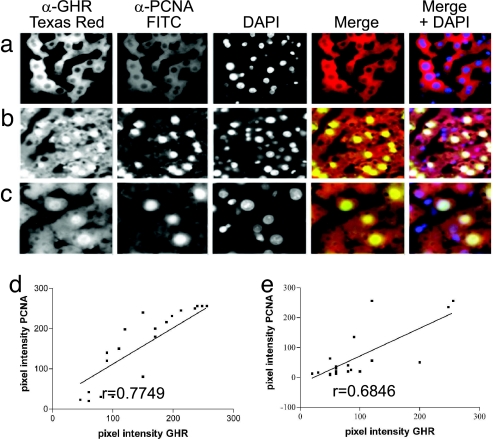
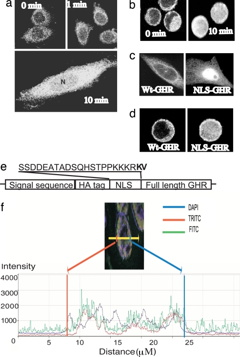
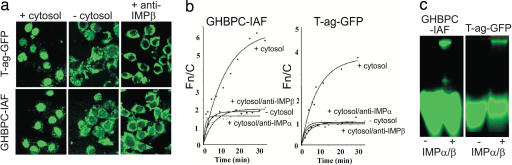
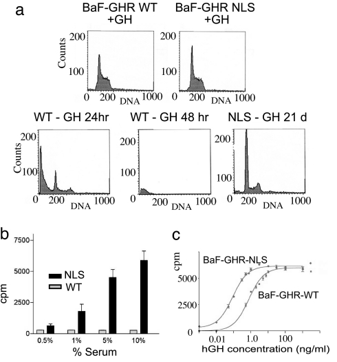
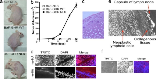
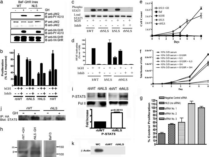
Growth hormone receptor (GHR) has been demonstrated to be nuclear localized both in vivo and in vitro, but the significance of this observation has remained elusive. Here we show that nuclear GHR is strongly correlated with proliferative status in vivo by using a liver regeneration model. In vitro, nuclear translocation of the GH receptor is GH-dependent and appears to be mediated by the Importin system. Constitutive nuclear targeting of GHR in murine pro-B cells is associated with constitutive activation of STAT5, a transforming agent in lymphoma and other cell types. This activation is abrogated by inhibition of JAK2 and appears to be driven by autocrine murine GH action coupled with enhanced nuclear uptake of phospho-STAT5. Nuclear targeting induces dysregulated cell cycle progression in the pro-B cell line, associated with constitutive up-regulation of the proliferation inducers Survivin and Mybbp, the metastasis related Dysadherin, and other tumor markers. GHR nuclear-targeted cells generate aggressive metastatic tumors when injected into nude mice, which display nuclear localized GHR strikingly similar to that seen in human lymphomas. We conclude that aberrant nuclear localization of GHR is a marker of high proliferative status and is sufficient to induce tumorigenesis and tumor progression.


Conflict of interest statement

The authors declare no conflict of interest.

Figures

Fig. 1.
Fig. 1.
Nuclear localization of GHR in regenerating rat liver. (ac) Liver from a control animal (a) and 24 h after hepatectomy (b and c) is shown. GHR localization was detected with MAb 263 (left image) and proliferation as PCNA with mAb PC10-FITC (second image). Nuclei were stained with DAPI (third image). The merged image in the fourth image is for GHR and PCNA immunoreactivity. Blue nuclei in Right represent DAPI staining, whereas white nuclei colocalize all three. Higher magnification (×60) shows more detail in regenerating liver 24 h after hepatectomy (c). Negative controls for staining specificity where primary antibody (mAb 263) was omitted showed no staining (not shown). (d and e) Correlation plots show the correlation between fluorescence intensity for mAb263 (GHR) and mAb PC10 (PCNA) in the nuclei of randomly chosen hepatocytes at 24 h after confocal scanning light microscopy in Hx (d) and sham Hx (e) livers.
Fig. 2.
Fig. 2.
Nuclear translocation of GHR occurs in response to GH. Immunofluorescence/CLSM imaging shows rapid GH-dependent nuclear translocation of GHR in CHO-K1 and BaF stable lines. GHR was detected by mAb 263 and FITC-coupled secondary antibody. (a) After 10-h serum starve, nuclear immunofluorescence was absent in CHO-GHR cells. (b) Addition of GH (100 ng/ml, 5 nM) resulted in rapid accumulation of GHR around the nuclear membrane at 1 min, followed by nuclear localization, which reaches a maximum at 10-min CLSM with anti-HA for HA-tagged GHR also shows rapid GH-dependent nuclear translocation of GHR in BaF-GHR WT stable lines. (c and d) Addition of the T-Ag NLS to the N terminus of mature GHR resulted in constitutive nuclear localization of GHR in the absence of GH, in both CHO-K1 (c) and BaF (d) cells. (e) Location and sequence of NLS are shown in e. (f) Quantification of 4.1 μm confocal scanning light microscopy z-scan of NLS-GHR transiently transfected COS cell, scan across one cell. Nucleus is DAPI stain (blue line), HA tag (N terminus, FITC, green) colocalizes with FLAG tag (C terminus, TRITC, red), quantified by scan across nucleus.
Fig. 3.
Fig. 3.
Nuclear accumulation of GHR in vitro is mediated by a classical nuclear import pathway involving Impα and -β. (a) CLSM visualization showing localization of T-Ag-GFP (Upper) and GHBPC (GHBPC-IAF) (Lower) in mechanically perforated HTC rat hepatoma cells in the absence or presence of cytosol or cytosol with anti-IMPβ antibody. (b) Quantitative measurements of the kinetics of nuclear accumulation of GHBPC (Left) and T-Ag-GFP (Right), +/− exogenous cytosol in the absence or presence of specific antibodies to IMPα or IMPβ. [Data were fitted for the function Fn/c(t) = Fn/cmax × (1− e-kt], where t is time, Fn/cmax is the maximal level of nuclear accumulation, and k is the first-order rate constant. (c) Recognition of GHBPC by the IMP α/β heterodimer as indicated by native gel electrophoresis imaging; 25 pmol GHBPC-IAF or T-Ag-GFP was incubated in the absence or presence of 10 μM IMPα/β heterodimer for 15 min at 20°C, before electrophoresis.
Fig. 4.
Fig. 4.
Effect of increased GHR nuclear targeting in a cell model. In a subconfluent culture with GH support, both BaF-GHR WT and NLS lines undergo mitosis, with 60% in G1 phase and 40% in S/M phase (a Upper). However, upon the withdrawal of cytokine support (in the form of GH or IL-3), only the BaF-GHR NLS lines proliferate (a Lower; only 21-day sort shown for NLS line, exemplifying earlier timepoints). (b) BaF-GHR NLS lines continue to proliferate in the absence of GH and IL-3, with the support of fetal bovine serum. (c) NLS-GHR lines show a dramatic increase in the sensitivity to GH as measured by [3H]thymidine incorporation assay (20-fold decrease in ED50, i.e., 10 pM for NLS, compared with 200 pM for WT in 1.0% serum). Results were confirmed in three separate population studies and clonal lines from three separate transfections.
Fig. 5.
Fig. 5.
s.c. inoculation of BaF-GHR NLS cells results in rapid tumor formation in vivo. (a) BaF-GHR NLS cells give rise to aggressive metastatic tumors when injected s.c. into nude mice. (b) Quantitative data over a 16-day period are shown graphically. (d Upper) No tumors were observed in the BaF-GHR WT or the BaF-NLS (as a negative control for the NLS sequence) group. HA-GHR immunofluorescence by using TRITC second antibody shows the tumors are derived from BaF HA-GHR NLS cells. (d Lower) A negative control omitting the primary anti-HA antibody shows staining specificity. (c) s.c. tumor samples and lymph nodes were taken from all BaF GHR NLS mice, as well as a liver sample from one of the group, and stained by H&E. (e) Nuclear localization of GHR in human lymphoma. (f) Negative control for staining in human lymphoma where primary antibody (mAb 263) is omitted.
Fig. 6.
Fig. 6.
Mechanistic studies. (a) Activation of WT and NLS GHR in BaF lines as determined by tyrosine phosphorylation of JAK2 and GHR in phosphotyrosine immunoblots. BaF-GHR WT or NLS lines were serum-starved for 6 h and then treated with human GH (100 ng/ml) (+) or saline (−) for 10 min. (b) Proliferative response of BaF lines, and abrogation of this with JAK2 inhibitor 1 (Calbiochem, San Diego, CA) at 0.4 μM, using the 3-(4,5-dimethylthiazol-2-yl)-2,5-diphenyl tetrazolium bromide assay (MTT) (SI Text). (c) STAT5 activation in BaF lines showing constitutive activation in BaF-NLS and abrogation by JAK inhibitor, with quantification for three independent experiments (d). (e) Proliferation of BaF lines from 0.5 × 10⁶ cells showing lack of constitutive proliferation with NLS-human GHR cells. (f) G120R (0.6 μM) blocks constitutive proliferation of NLS-rbGHR expressing cells growing in charcoal stripped serum (g) siRNA to murine GH inhibits constitutive proliferation in charcoal stripped serum (h) mature and precursor full-length GHR in highly purified BaF cell nuclei prepared according to (28). (i) Enhanced basal (2.5-fold by densitometry) phospho-STAT5 (Cell Signaling clone 14H2, Y694) in purified nuclei of NLS-rbGHR BaFcells. Histogram shows quantification of three separate preparations, mean ± SEM (j) STAT5 coimmunoprecipitation with nuclear GHR, even in the basal state with NLS-GHR BaF cells. (k) β-Actin immunoblot comparing whole-cell and nuclear β-actin with 10 μg of protein, demonstrating purity of nuclei in i.

References

    1. Lobie PE, Barnard R, Waters MJ. J Biol Chem. 1991;266:22645–22652. - PubMed
    1. Mertani HC, Raccurt M, Abbate A, Kindblom J, Tornell J, Billestrup N, Usson Y, Morel G, Lobie PE. Endocrinology. 2003;144:3182–3195. - PubMed
    1. Lobie PE, Wood TJ, Chen CM, Waters MJ, Norstedt G. J Biol Chem. 1994;269:31735–31746. - PubMed
    1. Lin SY, Makino K, Xia W, Matin A, Wen Y, Kwong KY, Bourguignon L, Hung MC. Nat Cell Biol. 2001;3:802–808. - PubMed
    1. Johnson HM, Subramaniam PS, Olsnes S, Jans DA. BioEssays. 2004;26:993–1004. - PubMed

Publication types

Associated data