. 2007 Nov 15;110(10):3673-81.
doi: 10.1182/blood-2007-04-087171. Epub 2007 Aug 9.

Treatment with GITR agonistic antibody corrects adaptive immune dysfunction in sepsis

Affiliations

Treatment with GITR agonistic antibody corrects adaptive immune dysfunction in sepsis

Philip O Scumpia et al. Blood. .

Abstract

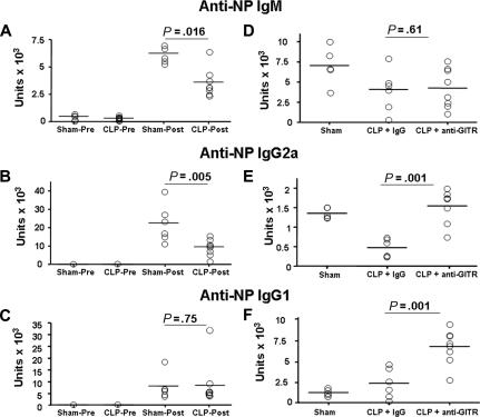
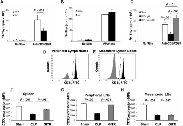
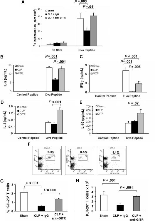
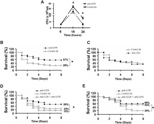
Apoptosis of CD4(+) T cells and T(H)2 polarization are hallmarks of sepsis-induced immunoparalysis. In this study, we characterized sepsis-induced adaptive immune dysfunction and examined whether improving T-cell effector function can improve outcome to sepsis. We found that septic mice produced less antigen-specific T-cell-dependent IgM and IgG(2a) antibodies than sham-treated mice. As early as 24 hours after sepsis, CD4(+) T cells proliferated poorly to T-cell receptor stimulation, despite normal responses to phorbol myristate acetate and ionomycin, and possessed decreased levels of CD3zeta. Five days following immunization, CD4(+) T cells from septic mice displayed decreased antigen-specific proliferation and production of IL-2 and IFN-gamma but showed no difference in IL-4, IL-5, or IL-10 production. Treatment of mice with anti-GITR agonistic antibody restored CD4(+) T-cell proliferation, increased T(H)1 and T(H)2 cytokine production, partially prevented CD3zeta down-regulation, decreased bacteremia, and increased sepsis survival. Depletion of CD4(+) T cells but not CD25(+) regulatory T cells eliminated the survival benefit of anti-GITR treatment. These results indicate that CD4(+) T-cell dysfunction is a key component of sepsis and that improving T-cell effector function may be protective against sepsis-associated immunoparalysis.


Figures

Figure 1
Figure 1
Anti-GITR treatment improves T-cell–dependent antibody responses in septic mice. C57Bl/6 mice underwent a sham procedure or cecal ligation and puncture (CLP) surgery and were immunized subcutaneously with the T-cell–dependent antigen NP-KLH and alum. Ten days later, serum (A) IgM, (B) IgG2a, and (C) IgG1 anti-NP antibody responses were measured by ELISA and compared with baseline samples. In another experiment, C57Bl/6 mice were given an intraperitoneal injection of 300 μg control antibody (Sham and CLP groups) or anti-GITR antibody and underwent cecal ligation and puncture or sham treatment 30 minutes later. Immediately following, mice were immunized with NP-KLH and alum. Ten days later, mice were bled and anti-NP–specific IgM (A), IgG2a (B), and IgG1 (C) were measured from the serum. P values indicate the difference between groups after immunization by Student t test (A-C) or post hoc analysis using Fisher LSD method (D-F).
Figure 2
Figure 2
T cells from septic mice proliferate poorly to TCR stimulation and display decreased CD3ζ expression. Twenty-four hours after cecal ligation and puncture (CLP) or sham treatment, CD25-depleted CD4+ T-effector cells were harvested as described in “Proliferation assay.” T-effector cells (2.5 × 10⁴) were cultured for 72 hours with anti-CD3 (2.5 μg/mL) and anti-CD28 (1 μg/mL; A) or PMA and ionomycin (B). 3H-thymidine was added during the last 14 to 18 hours of culture and incorporation was measured using a liquid scintillation counter. (C) Mice were treated as in panel A, except prior to CLP, mice were given an intraperitoneal injection of anti-GITR or control antibody (300 μg). Mice were given 300 μg anti-GITR or control antibody at the time of CLP surgery. Twenty-four hours after CLP or sham surgery, cells were stained extracellularly for flow cytometry using anti-CD3ϵ Pacific Blue and anti-CD4 allophycocyanin. Cells were then fixed and permeabilized as described in “Flow cytometry” and stained for anti-CD3ζ FITC. (D,E) Histogram overlays showing CD3ζ from CD4+ T cells of 1 sham mouse (filled black) versus 1 control antibody–treated CLP mouse (solid black line, no fill) versus 1 anti-GITR–treated CLP mouse (dotted line, no fill) and an isotype control antibody (filled gray) from peripheral lymph nodes (D) or mesenteric lymph nodes (E). The calculated mean fluorescent intensity (MFI) from (F) spleen, (G) peripheral lymph nodes, and (H) mesenteric lymph nodes from n = 3 mice per group. Histogram overlays were made using FCS Express software version 3 (De Novo, Los Angeles, CA). P values are indicative of the difference between groups using Student t test (A) or post hoc analysis using the Fisher LSD method. All error bars indicate SD.
Figure 3
Figure 3
Sepsis-induced antigen-specific CD4+ T-cell dysfunction is corrected by anti-GITR treatment. DO11.10 mice were administered 300 μg anti-GITR or control antibody and were immunized 30 minutes later with Ova323-339 in alum at the time of cecal ligation and puncture or sham surgery. Five days later, lymph node cells were harvested and 2.5 × 10⁴ CD4+ cells were cultured with 2.5 × 10⁵ irradiated APCs with Ova peptide for 72 hours. (A) Proliferation of CD4+ T cells was measured as the incorporation of 3H-thymidine during the last 18 hours of culture. Media was harvested at the time of 3H-thymidine addition to assess cytokine production. (B) IL-2, (C) IFN-γ, (D) IL-4, and (E) IL-10 concentrations were assessed by Luminex multiplex analysis. (F) Representative examples of flow plots demonstrating the expansion of DO11.10 T cells (KJI-26+CD4+) from lymph nodes of Balb/c mice that were injected intravenously with 5 × 10⁶ DO11.10 CD4+ T cells 3 days before sham (left), CLP with control antibody (middle), or CLP with anti-GITR antibody (right) treatment at the time of immunization with Ova323-337 peptide in alum. (G) The calculated percentage of living (Sytox Blue), nondebris DO11.10 T cells (KJI26+CD4+) expanded in the peripheral lymph nodes of the Balb/c mice. (H) The calculated total number of live (Sytox Blue), nondebris DO11.10 T cells using cell counts obtained with a hemacytometer. P values indicate differences between groups after post hoc analysis using the Fisher LSD method.
Figure 4
Figure 4
Anti-GITR treatment decreases bacteremia and improves sepsis survival. (A) C57Bl/6 mice were given an intraperitoneal injection of 300 μg anti-GITR (n = 9) or control antibody (n = 9) 30 minutes before cecal ligation and puncture (CLP) surgery and mice were killed at 6, 16, or 24 hours afterward. Bacteremia was determined from blood obtained aseptically via cardiac puncture plated on sheep blood agar plates. Data are represented as mean ± standard error. (B) C57Bl/6 mice were given an intraperitoneal injection of 300 μg anti-GITR (n = 35) or control antibody (n = 38) 30 minutes before CLP surgery and survival was monitored for 8 days. Figure is the combination of 2 separate experiments with similar results. (C) C57Bl/6 mice were given injections of CD4 depleting (n = 15) or control antibody (n = 20) as described in “Proliferation assay.” Survival after CLP was monitored for 8 days. (D) C57Bl/6 mice depleted of CD4 cells (n = 20) or not (n = 20) were given anti-GITR antibody 30 minutes before CLP surgery. Another group of non-CD4–depleted mice receiving control antibody was used as a control group. (E) C57Bl/6 mice depleted of CD25+ cells (n = 20) or not (n = 20) were given anti-GITR antibody 30 minutes before CLP surgery. Another group of non-CD25–depleted mice receiving control antibody was used as a control group.*P < .05 by Student t test (A) or Fisher exact test (B-E). All error bars indicate SD.

References

    1. Hotchkiss RS, Chang KC, Swanson PE, et al. Caspase inhibitors improve survival in sepsis: a critical role of the lymphocyte. Nat Immunol. 2000;1:496–501. - PubMed
    1. Hotchkiss RS, Tinsley KW, Swanson PE, et al. Prevention of lymphocyte cell death in sepsis improves survival in mice. Proc Natl Acad Sci U S A. 1999;96:14541–14546. - PMC - PubMed
    1. Wesche-Soldato DE, Chung CS, Lomas-Neira J, Doughty LA, Gregory SH, Ayala A. In vivo delivery of caspase-8 or Fas siRNA improves the survival of septic mice. Blood. 2005;106:2295–2301. - PMC - PubMed
    1. Hotchkiss RS, Swanson PE, Knudson CM, et al. Overexpression of Bcl-2 in transgenic mice decreases apoptosis and improves survival in sepsis. J Immunol. 1999;162:4148–4156. - PubMed
    1. Docke WD, Randow F, Syrbe U, et al. Monocyte deactivation in septic patients: restoration by IFN--gamma treatment. Nat Med. 1997;3:678–681. - PubMed

Publication types

MeSH terms

Substances