. 2007 Dec 1;110(12):4077-85.
doi: 10.1182/blood-2007-02-073841. Epub 2007 Aug 9.

Kupffer cell heterogeneity: functional properties of bone marrow derived and sessile hepatic macrophages

Affiliations

Kupffer cell heterogeneity: functional properties of bone marrow derived and sessile hepatic macrophages

Ingo Klein et al. Blood. .

Abstract

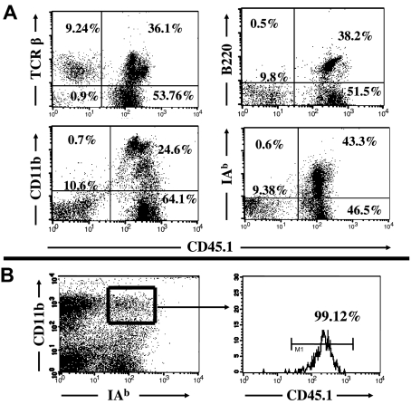
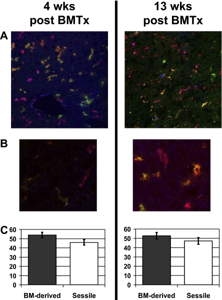
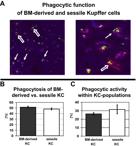
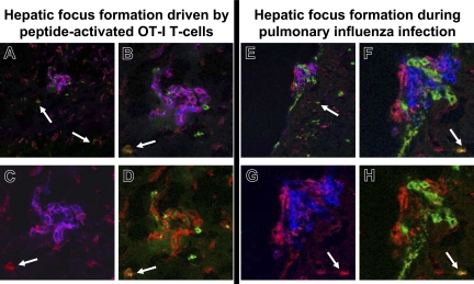
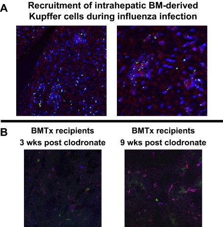
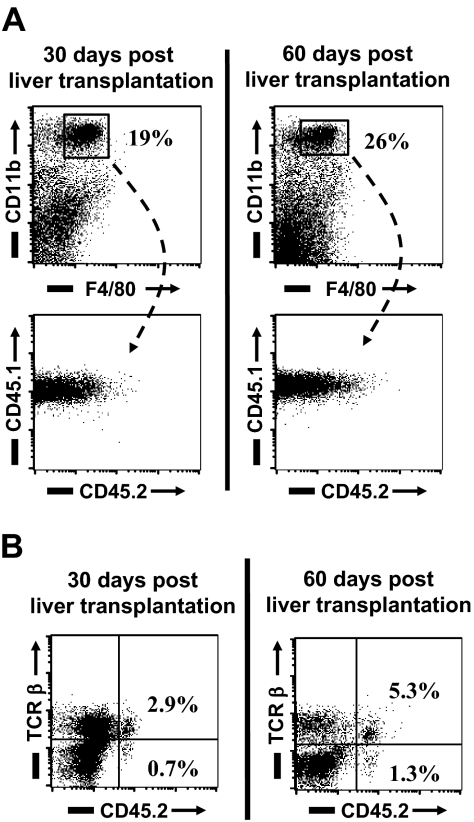
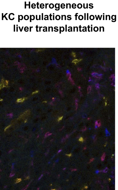
Kupffer cells form a large intravascular macrophage bed in the liver sinusoids. The differentiation history and diversity of Kupffer cells is disputed; some studies argue that they are derived from blood monocytes, whereas others support a local origin from intrahepatic precursor cells. In the present study, we used both flow cytometry and immunohistochemistry to distinguish 2 subsets of Kupffer cells that were revealed in the context both of bone marrow transplantation and of orthotopic liver transplantation. One subset was radiosensitive and rapidly replaced from hematogenous precursors, whereas the other was relatively radioresistant and long-lived. Both were phagocytic but only the former population was recruited into inflammatory foci in response to CD8(+) T-cell activation. We propose the name "sessile" for the radioresistant Kupffer cells that do not participate in immunoinflammatory reactions. However, we found no evidence that these sessile Kupffer cells arise from immature intrahepatic precursors. Our conclusions resolve a long-standing controversy and explain how different experimental approaches may reveal one or both of these subsets.


Figures

Figure 1
Figure 1
Flow-cytometric analysis of recipient leukocyte replacement in radiation bone marrow chimeras. The replacement of various leukocyte populations in B6.CD45.2 mice was analyzed 4 weeks after 10-Gy radiation and congenic bone marrow transplantation from B6.CD45.1 bone marrow donors. (A) Approximately 90% of intrahepatic leukocytes were replaced by donor-type leukocytes. The remaining 10% were almost entirely TCR-positive (top left) and negative for CD45R/B220, CD11b, and MHC class II, indicating that they were non-Kupffer cells. (B) The origin of isolated intrahepatic macrophages was greater than 99% donor BM-derived, based on expression of CD11b and MHC class II (IAb). Numbers on plots are percentages of lymphocytes.
Figure 2
Figure 2
Immunohistologic staining for Kupffer cells in radiation bone marrow chimeras. B6.CD45.2 recipient mice were irradiated (10 Gy) and reconstituted with B6.CD45.1 bone marrow. Frozen tissue sections were stained for macrophages (F4/80), CD45.1+ BM-derived cells, and CD45.2+ recipient-derived cells, revealing Kupffer cells derived from donor BM (purple-red), sessile recipient-type KCs (orange-green), BM-derived non-KC leukocytes (blue), and recipient-type non-KC leukocytes (green). (A) Liver tissue sections 4 weeks (left) and 13 weeks (right) after bone marrow transplantation, showing an equal distribution of BM-derived and sessile KCs. Original magnification, ×200. (B) Higher magnification of BM-derived and sessile KCs 4 weeks (left) and 13 weeks (right) after bone marrow transplantation. Original magnification, ×400. (C) Quantitative analysis of the 2 KC subsets revealed that 46% (± 3%) were sessile KCs of recipient origin. Error bars represent SEM.
Figure 3
Figure 3
Sessile and BM-derived KCs have equal phagocytic capacity. To evaluate the phagocytic capacity of BM-derived and sessile KCs, radiation bone marrow chimeras (10-Gy radiation dose) were injected with fluorescent beads, which underwent rapid phagocytosis by macrophages. (A, left) The distribution of fluorescent beads in BM-derived and sessile KCs was assessed by costaining with a fluorescent anti-CD45.1 mAb. Double-positive CD45.1 KCs appeared purple (closed arrow), whereas sessile (CD45.2+) KCs only stained with F4/80 (red, open arrow). Original magnification, ×200 (left). Higher magnification, ×400 (right). (B) Quantitative analysis of bead-carrying KCs revealed an almost equal distribution of phagocytic BM-derived and sessile KCs (52% ± 1.7% vs 48% ± 1.7%). (C) Phagocytic activity of BM-derived (26% ± 1.7%) versus sessile KCs (32% ± 5.3%) expressed as a fraction of the total KCs.
Figure 4
Figure 4
Only BM-derived KCs participate in focal intrahepatic inflammation. Liver sections of bone marrow transplant recipients were stained with CD45.2-FITC (green), CD45.1-Cy5 (displayed in false color blue), and F4/80-PE (red). Inflammatory foci developed during peptide-activated intrahepatic accumulation of adoptively transferred TCR-tg OT-I cells (A-D) or with accumulation of endogenous CD8+ T cells during pulmonary influenza infection (E-H). (A,E) In both models, sessile KCs (orange, closed arrow) were only detectable outside the foci. Kupffer cells within the foci are all BM derived (purple) with multiple BM-derived and recipient-type non-KC leukocytes within the foci (blue and green). Original magnification, ×200. (B,F) Enlarged (×400) inflammatory focus following adoptive OT-I transfer and peptide activation (B) or during pulmonary influenza infection (F). (C,D,G,H) Two-color reproductions of the same foci with F4/80-CD45.1 costaining for BM-derived KCs (C,G) and F4/80-CD45.2 costaining for sessile KCs (D,H) confirmed the absence of sessile KCs within inflammatory foci.
Figure 5
Figure 5
Inflammatory BM-derived KCs were not directly recruited from the bone marrow. (A) Bone marrow chimeras were injected with fluorescent microspheres and then infected with influenza. Microspheres were immediately removed from the circulation by phagocytes, but focus formation within the livers did not occur until 5 days after infection. The accumulation of bead-carrying KCs indicated that KCs engaging in focus formation had already populated the liver several days earlier and were not derived from circulating monocytes. KCs (F4/80-Cy5) are displayed in false color blue, DAPI-stained nuclei are red, and fluorescent microspheres green. (B) To evaluate if the sessile KC population was derived from radioresistant intrahepatic precursor cells, hepatic KCs were entirely eliminated by clodronate liposome injection 3 weeks after bone marrow transplantation. Three weeks or 9 weeks later, sections from bone marrow chimeras were stained for BM-derived (CD45.1, blue) and sessile (CD45.2, green) KCs (F4/80, red). While recipient CD45.2 non-KC leukocytes (lymphocytes, granulocytes, etc) were abundant 3 and 9 weeks after clodronate injection, there were only BM-derived KCs in the liver sections of these animals; sessile KCs were not detectable. Original magnification, ×400.
Figure 6
Figure 6
Flow-cytometric analysis of recipient leukocyte replacement in orthotopic liver transplant recipients. As an alternative approach to the analysis of KC turnover, livers from B6.CD45.2 animals were transplanted orthotopically into B6.CD45.1 recipients. By this experimental design, the bone marrow compartment of the recipient was spared from experimental manipulation. (A) The replacement of CD45.2 intrahepatic leukocytes by CD45.1 BM-derived cells was evaluated 30 and 60 days after liver transplantation by flow-cytometric analysis. Similar to bone marrow chimeras, CD11b/F4/80-positive KCs were all CD45.1+ in cell isolates from intrahepatic leukocytes. This indicated their origin from the bone marrow of the liver transplant recipient. (B) As seen in radiation bone marrow chimeras, a small percentage of CD45.2/TCR+ cells remained within the transplanted livers. Numbers in plots are percentages of lymphocytes.
Figure 7
Figure 7
Immunohistologic staining of liver transplants reveals equal numbers of BM-derived and sessile KCs. Frozen tissue sections from liver transplants (B6.CD45.2 liver transplanted into B6.CD45.1 recipient) were stained for macrophages (F4/80 PE), CD45.1 Cy5 (displayed in false color blue) BM-derived cells, and CD45.2 FITC (green) liver transplant-derived cells. Equal numbers of BM-derived ‘KCs (purple-red) and sessile KCs (orange-green) were detected by confocal microscopy.

References

    1. Bayon LG, Izquierdo MA, Sirovich I, van Rooijen N, Beelen RH, Meijer S. Role of Kupffer cells in arresting circulating tumor cells and controlling metastatic growth in the liver. Hepatology. 1996;23:1224–1231. - PubMed
    1. Lehner PJ, Cresswell P. Recent developments in MHC-class-I-mediated antigen presentation. Curr Opin Immunol. 2004;16:82–89. - PubMed
    1. Rogoff TM, Lipsky PE. Antigen presentation by isolated guinea pig Kupffer cells. J Immunol. 1980;124:1740–1744. - PubMed
    1. Seki S, Habu Y, Kawamura T, et al. The liver as a crucial organ in the first line of host defense: the roles of Kupffer cells, natural killer (NK) cells and NK1. 1 Ag+ T cells in T helper 1 immune responses. Immunol Rev. 2000;174:35–46. - PubMed
    1. Wiegard C, Frenzel C, Herkel J, Kallen KJ, Schmitt E, Lohse AW. Murine liver antigen presenting cells control suppressor activity of CD4+CD25+ regulatory T cells. Hepatology. 2005;42:193–199. - PubMed

Publication types

MeSH terms