. 2007 Sep 5;26(17):3936-44.
doi: 10.1038/sj.emboj.7601817. Epub 2007 Aug 9.

Structural insights into the transcriptional and translational roles of Ebp1

Affiliations

Structural insights into the transcriptional and translational roles of Ebp1

Tom P Monie et al. EMBO J. .

Abstract

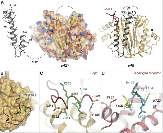
The ErbB3-binding protein 1 (Ebp1) is an important regulator of transcription, affecting eukaryotic cell growth, proliferation, differentiation and survival. Ebp1 can also affect translation and cooperates with the polypyrimidine tract-binding protein (PTB) to stimulate the activity of the internal ribosome entry site (IRES) of foot-and-mouth disease virus (FMDV). We report here the crystal structure of murine Ebp1 (p48 isoform), providing the first glimpse of the architecture of this versatile regulator. The structure reveals a core domain that is homologous to methionine aminopeptidases, coupled to a C-terminal extension that contains important motifs for binding proteins and RNA. It sheds new light on the conformational differences between the p42 and p48 isoforms of Ebp1, the disposition of the key protein-interacting motif ((354)LKALL(358)) and the RNA-binding activity of Ebp1. We show that the primary RNA-binding site is formed by a Lys-rich motif in the C terminus and mediates the interaction with the FMDV IRES. We also demonstrate a specific functional requirement for Ebp1 in FMDV IRES-directed translation that is independent of a direct interaction with PTB.


Figures

Figure 1
Figure 1
Molecular structure of Ebp1 and comparison with a type II human MAP (hMAP2). (A) Ribbon diagram of the crystal structure of Ebp1(8–360); α-helices are coloured pink and β-strands blue. (B) Superposition of Ebp1 in orange and hMAP2 in cyan (PDB 1kq9 (Nonato et al, 2006)); insertions in Ebp1 are coloured dark red and those in hMAP2 dark blue. (CE) Comparison of the active site of hMAP2 with the corresponding region in Ebp1. Side chains of selected residues are shown as sticks for (C) hMAP2, (D) a superposition of hMAP2 and Ebp1 and (E) Ebp1. The colour coding is the same as for panel B.
Figure 2
Figure 2
The C-terminal Lys-rich motif of Ebp1 binds the FMDV IRES RNA. EMSAs were performed using 32P-labelled FMDV IRES RNA (nt 280–716) and twofold dilutions of various Ebp1 proteins over the range 0.125–8 μM. (A) Ebp1(1–394); (B) Ebp1(1–394LM); (C) Ebp1(1–375) and (D) Ebp1(1–360). Ebp1 binds multiple regions of the FMDV IRES. EMSAs were performed using 32P-labelled RNA and twofold dilutions of full-length Ebp1 (residues 1–394) over the range 0.125–32 μM. (E) FMDV IRES Domain H (nts 280–336); (F) Domain I (nts 337–551) and (G) Domain JKL (nts 552–716). (H) Schematic secondary structure of the FMDV IRES.
Figure 3
Figure 3
Ebp1 interacts with PTB in an RNA-dependent manner. (A) 35S Methionine labelled coupled transcription-translation products for Ebp1 and control constructs. (B) GST pull-down assays between 35S-labelled Ebp1 constructs and 35 picomoles of either GST-Sxl (top panel) or GST-PTB (bottom panel). Reactions were performed in the absence (−) and presence (+) of RNAse treatment.
Figure 4
Figure 4
Ebp1 is required for efficient translation from the FMDV IRES. (A) Schematic representation of reporter RNAs used to analyse the effect of Ebp1-specific siRNAs on IRES-mediated translation. (B) Western blot analysis of cells transfected with either EGFP or Ebp1-specific siRNAs. Blots were probed with antisera to Ebp1, PTB or GAPDH. (C) Levels of the reporter proteins CAT and luciferase produced in cells transfected with either EGFP- or Ebp1-specific siRNAs. Levels are expressed as a percentage of that observed in the EGFP siRNA-transfected control. Assays were carried out in triplicate and the error bars represent the observed standard deviation.
Figure 5
Figure 5
Structural features at the N and C termini of Ebp1. (A) The structure shows that the predicted p42 isoform (left) which starts at Met 55 lacks one and a half helices at the N terminus of the p48 isoform (indicated in grey in the structure on the right). This helix makes extensive hydrophobic contacts with the body of Ebp1 (coloured by atom type: carbon—orange; nitrogen—blue oxygen—red; sulphur—yellow); its removal exposes a large hydrophobic cleft on one face of the protein. The structure of p48 Ebp1 also illustrates the proximity of K20 and K22 to the lys-rich loop 1; together these features may constitute a bipartite nucleolar localisation signal (Squatrito et al, 2004; Fujiwara et al, 2006). (B) Position of the 354LKALL358 protein-binding motif at the C terminus of Ebp1. Colouring is the same as in Figure 1B except that residues from the motifs are highlighted in green. The surface of Ebp1 up to residue 337 is shown. Close-up views (in similar orientations) of the LxxLL motif from (C) Ebp1 and (D) the AR ((Hur et al, 2004); PDB—1t7f). Residues from Ebp1 are colour coded as described above. Carbon atoms of the LxxLL motif of the peptide ligand of AR are cyan.

Similar articles

Cited by

References

    1. Ahn JY, Liu X, Liu Z, Pereira L, Cheng D, Peng J, Wade PA, Hamburger AW, Ye K (2006) Nuclear Akt associates with PKC-phosphorylated Ebp1, preventing DNA fragmentation by inhibition of caspase-activated DNase. EMBO J 25: 2083–2095 - PMC - PubMed
    1. Bazan JF, Weaver LH, Roderick SL, Huber R, Matthews BW (1994) Sequence and structure comparison suggest that methionine aminopeptidase, prolidase, aminopeptidase P, and creatinase share a common fold. Proc Natl Acad Sci USA 91: 2473–2477 - PMC - PubMed
    1. Bose SK, Sengupta TK, Bandyopadhyay S, Spicer EK (2006) Identification of Ebp1 as a component of cytoplasmic bcl-2 mRNP (messenger ribonucleoprotein particle) complexes. Biochem J 396: 99–107 - PMC - PubMed
    1. Brunger AT, Adams PD, Clore GM, DeLano WL, Gros P, Grosse-Kunstleve RW, Jiang JS, Kuszewski J, Nilges M, Pannu NS, Read RJ, Rice LM, Simonson T, Warren GL (1998) Crystallography and NMR system: a new software suite for macromolecular structure determination. Acta Crystallogr D 54: 905–921 - PubMed
    1. Collaborative Computer Project No. 4 (1994) The CCP4 suite: programs for protein crystallography. Acta Crystallogr D 50: 760–763 - PubMed

Publication types

MeSH terms