. 2007 Sep 10;313(15):3261-75.
doi: 10.1016/j.yexcr.2007.07.009. Epub 2007 Jul 19.

The stimulus-dependent co-localization of serum- and glucocorticoid-regulated protein kinase (Sgk) and Erk/MAPK in mammary tumor cells involves the mutual interaction with the importin-alpha nuclear import protein

Affiliations

The stimulus-dependent co-localization of serum- and glucocorticoid-regulated protein kinase (Sgk) and Erk/MAPK in mammary tumor cells involves the mutual interaction with the importin-alpha nuclear import protein

Patricia Buse et al. Exp Cell Res. .

Abstract

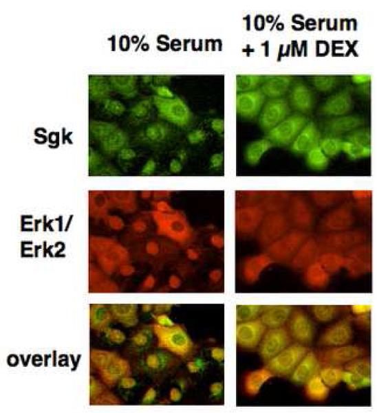
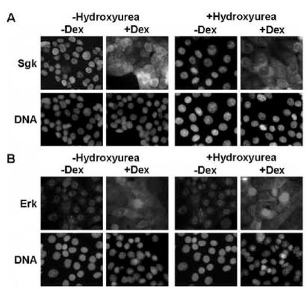
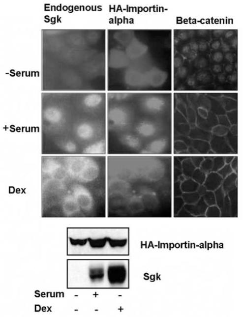
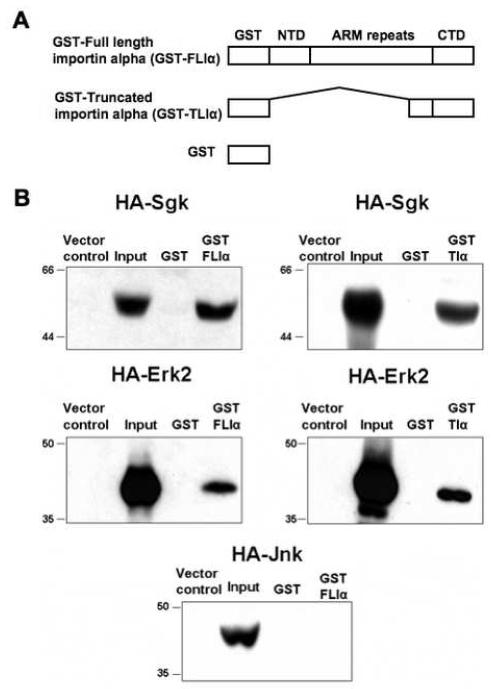
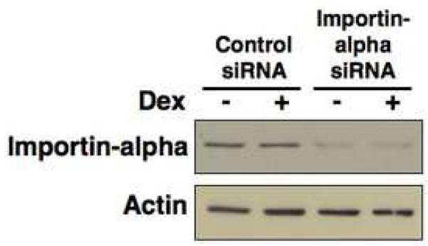
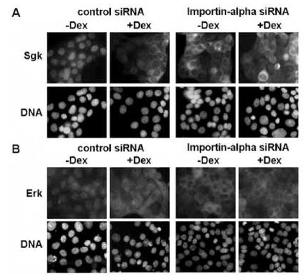
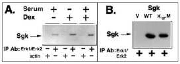
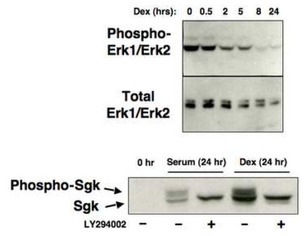
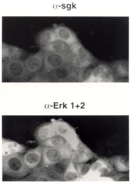
In Con8 rat mammary epithelial tumor cells, indirect immunofluorescence revealed that Sgk (serum- and glucocorticoid-regulated kinase) and Erk/MAPK (extracellular signal-regulated protein kinase/mitogen activated protein kinase) co-localized to the nucleus in serum-treated cells and to the cytoplasmic compartment in cells treated with the synthetic glucocorticoid dexamethasone. Moreover, the subcellular distribution of the importin-alpha nuclear transport protein was similarly regulated in a signal-dependent manner. In vitro GST-pull down assays revealed the direct interaction of importin-alpha with either Sgk or Erk/MAPK, while RNA interference knockdown of importin-alpha expression disrupted the localization of both Sgk and Erk into the nucleus of serum-treated cells. Wild type or kinase dead forms of Sgk co-immunoprecipitated with Erk/MAPK from either serum- or dexamethasone-treated mammary tumor cells, suggesting the existence of a protein complex containing both kinases. In serum-treated cells, nucleus residing Sgk and Erk/MAPK were both hyperphosphorylated, indicative of their active states, whereas, in dexamethasone-treated cells Erk/MAPK, but not Sgk, was in its inactive hypophosphorylated state. Treatment with a MEK inhibitor, which inactivates Erk/MAPK, caused the relocalization of both Sgk and ERK to the cytoplasm. We therefore propose that the signal-dependent co-localization of Sgk and Erk/MAPK mediated by importin-alpha represents a new pathway of signal integration between steroid and serum/growth factor-regulated pathways.


Figures

Fig. 1
Fig. 1
Effects of serum and glucocorticoids on the co-localization of Sgk and Erk/MAPK. Con8 mammary tumor cells were treated with 10% serum in the presence or absence of 1μM dexamethasone for 24 hours. The subcellular distribution of Sgk was examined by indirect immunofluorescence microscopy using affinity-purified rabbit polyclonal antibodies to Sgk followed by FITC-conjugated goat anti-rabbit secondary antibodies (green fluorescent staining). Erk1/Erk2 were detected using monoclonal antibodies that specifically recognized both MAPK family members followed by a rhodamine-conjugated anti-mouse secondary antibody employed to selectively visualize this protein kinase (red fluorescent staining). The orange color in the lower panels show the overlap of the green FITC staining and the red rhodamine signal.
Fig. 2
Fig. 2
Effects of hydroxyurea on the signal-dependent colocalization of Sgk and Erk/MAPK. Con8 cells were treated with 10% serum in the presence or absence of 1 mM hydroxyurea for 24 hours to induce a cell cycle arrest prior to treatment with 1 μM dexamethasone (Dex) for 24 hours. Panel A: The subcellular distribution of Sgk was examined by indirect immunofluorescence microscopy using affinity-purified rabbit polyclonal antibodies to Sgk followed by FITC-conjugated goat anti-rabbit secondary antibodies. Panel B: Erk1/Erk2 were detected using monoclonal antibodies that specifically recognized both MAPK family members followed by a rhodamine-conjugated anti-rabbit secondary antibody employed to selectively visualize this protein kinase. DNA was stained to visualize the nucleus using DAPI.
Fig. 3
Fig. 3
Stimulus-dependent localization of importin-alpha and endogenous Sgk. Low confluent (30%) mammary epithelial cells, grown on 2 well lab-tek slides were transfected with expression vectors encoding full length importin-alpha (HA-FLIa) using lipofectamine. The cells were serum starved for 36 hours, and then maintained without serum (−serum) or treated with either 10% calf serum (+ serum) or with 1 μM dexamethasone (Dex) for 15 hours. The localization of HA-importin-alpha (middle panels) and of endogenous Sgk (left panels) and beta-catenin (right panels) was examined by indirect immunoflorescence microscopy as described in the Materials and Methods section. Protein expression of HA-importin-alpha and endogenous Sgk under each of treatment conditions used for localization studies was evaluated by immunoblotting with anti-HA or anti-Sgk antibodies respectively.
Fig. 4
Fig. 4
Interaction of ectopically expressed HA-Erk2 and HA-Sgk to recombinant full length and truncated importin-alpha. Panel A: GST-FLIa (full length importin-alpha) fusion protein is composed of a GST epitope tag, amino-terminal domain (NTD), Arm repeat containing domain, and a carboxy-terminal domain (CTD). GST-Tia (truncated importin-alpha) is composed of GST linked to the carboxy-terminal 106 amino acids (amino acids 423-529). GST alone was used as a negative control. Panel B: Hek-293 cells were transiently transfected with expression plasmids encoding either wild-type HA epitope tagged Erk2 (HA-Erk2) or Sgk (WT-HA-Sgk) or HA-Jnk or vector alone, and 48 hours post-transfection cell lysates were prepared as described in the Methods section. GST protein alone or GST-FLIa or GST-TIa was incubated with the indicated lysates containing exogenously expressed wild-type HA-tagged Sgk, or HA-Erk2, or HA-Jnk. The proteins bound to the Glutathione bead-bound GST or GST-fusion proteins were separated on SDS-PAGE and immunoblotted with anti-HA antibodies to detect bound proteins. Inputs denote 10% of the extract and lysates prepared from vector transfected samples served as controls as shown in each panel. Molecular weight markers are shown on the left side of each panel. Experiments were repeated three times with similar results.
Fig. 5
Fig. 5
Knockdown of importin-alpha using RNA interference. Con8 cells were reverse transfected with 150 nM siRNA specific to importin-alpha or 150 nM of nonspecific (control) siRNA at the time of plating. Cells were then treated with or without 1 uM dexamethasone for 24 hours. Cell lysates were separated on SDS-PAGE and immunoblotted with anti-importin-alpha or actin specific antibodies as described in the Materials and Methods section.
Fig. 6
Fig. 6
Effects of RNAi knock down of importin-alpha on the localization of Sgk and ERK. Con8 cells were reverse transfected with 150 nM siRNA specific to importin-alpha or 150 nM of nonspecific (control) siRNA at the time of plating. Cells cultured in 10% serum were then treated with or without 1 uM dexamethasone (Dex) for 24 hours. Panel A: The subcellular distribution of Sgk was examined by indirect immunofluorescence microscopy using affinity-purified rabbit polyclonal antibodies to Sgk followed by FITC-conjugated goat anti-rabbit secondary antibodies. Panel B: Erk1/Erk2 were detected using monoclonal antibodies that specifically recognized both MAPK family members followed by a rhodamine-conjugated anti-rabbit secondary antibody employed to selectively visualize this protein kinase. DNA was stained to visualize the nucleus using DAPI.
Fig. 7
Fig. 7
Co-immunoprecipitation of Sgk with Erk/MAPK. Panel A: Cells were transiently transfected with a wild-type Sgk expression vector and then treated with the indicated combinations of 10% serum and/or 1 μM dexamethasone for 24 hours. Cell extracts were immunoprecipitated with antibodies directed against either Erk1/Erk2 or to actin, the immunoprecipitated material electrophoretically fractionated and probed for the presence of Sgk. Panel B: Cells were transfected with a wild-type (WT) or the kinase dead (K127M) forms of Sgk or with an empty expression vector (V). Erk1/Erk2 was immunoprecipitated from the cell extracts, electrophoretically fractionated and western blots probed for the presence of Sgk.
Fig. 8
Fig. 8
Effects of serum and glucocorticoids on production of the hyperphosphorylated forms of Sgk and Erk/MAPK. Serum-treated cells were incubated with 1 μM dexamethasone and at the indicated time points, total cell extracts were electrophoretically fractionated. Western blots were probed for production of activated MAPK (top panel) or total Erk1/Erk2 (middle panel) using the appropriate antibodies. Cells were treated with 10% serum or 1 μM dexamethasone (Dex) in presence or absence of 50 μM LY294002, which inhibits PI 3-kinase activity, for 24 hours. A control culture was maintained in the absence of either stimuli (0 hr). Cell extracts were electrophoretically fractionated and probed for Sgk using affinity purified anti-Sgk antibodies. The hyperphosphorylated and hypophosphorylated Sgk species are designated with arrows.
Fig. 9
Fig. 9
Effects of the PD 98095 MEK inhibitor on the subcellular localization of Sgk and Erk/MAPK. Con8 cells were treated with 10% serum for 18 hours and 50 uM PD 98095 for the last 8 hours. The subcellular distribution of Sgk and Erk/MAPK was examined by indirect immunofluorescence microscopy using affinity-purified rabbit polyclonal antibodies to Sgk (upper panel) or monoclonal anti-Erk 1 and Erk 2 antibodies (lower panel) as the primary antibodies respectively. The secondary antibodies were FITC-conjugated goat anti-rabbit and rhodamine-conjugated goat-anit-mouse, respectively.

Similar articles

Cited by

References

    1. Beato M, Chavez S, Truss M. Transcriptional regulation by steroid hormones. Steroids. 1996;61:240–51. - PubMed
    1. Beato M, Sanchez-Pacheco A. Interaction of steroid hormone receptors with the transcription initiation complex. Endocr. Rev. 1996;17:587–609. - PubMed
    1. Dupont J, Le Roith D. Insulin-like growth factor 1 and oestradiol promote cell proliferation of MCF-7 breast cancer cells: new insights into their synergistic effects. Mol. Pathol. 2001;54:149–54. - PMC - PubMed
    1. Johnson GL, Vaillancourt RR. Sequential protein kinase reactions controlling cell growth and differentiation. Curr. Opin. Cell Biol. 1994;6:230–8. - PubMed
    1. Karin M. The regulation of AP-1 activity by mitogen-activated protein kinases. J. Biol. Chem. 1995;270:16483–6. - PubMed

Publication types

MeSH terms

Substances