. 2007 Sep 15;366(1):16-22.
doi: 10.1016/j.virol.2007.06.034. Epub 2007 Aug 9.

Heparan sulfate is a binding molecule but not a receptor for CEACAM1-independent infection of murine coronavirus

Affiliations

Heparan sulfate is a binding molecule but not a receptor for CEACAM1-independent infection of murine coronavirus

Rie Watanabe et al. Virology. .

Abstract

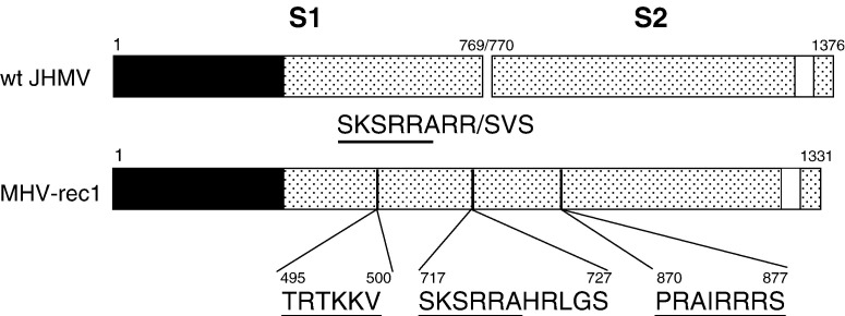
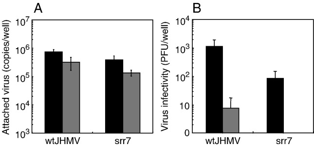
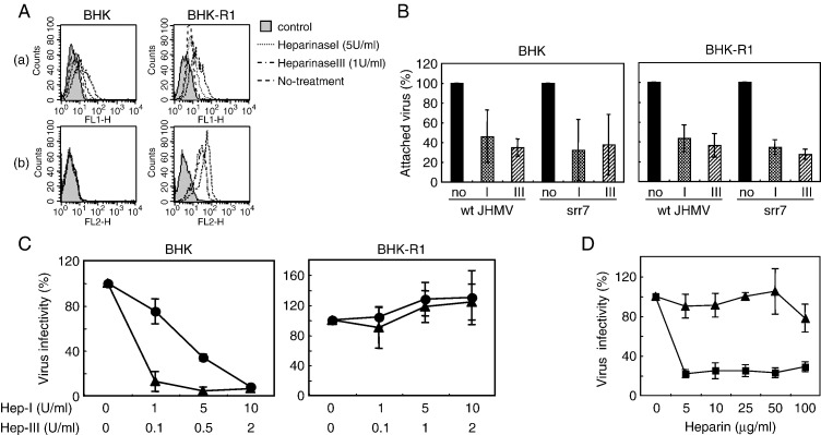
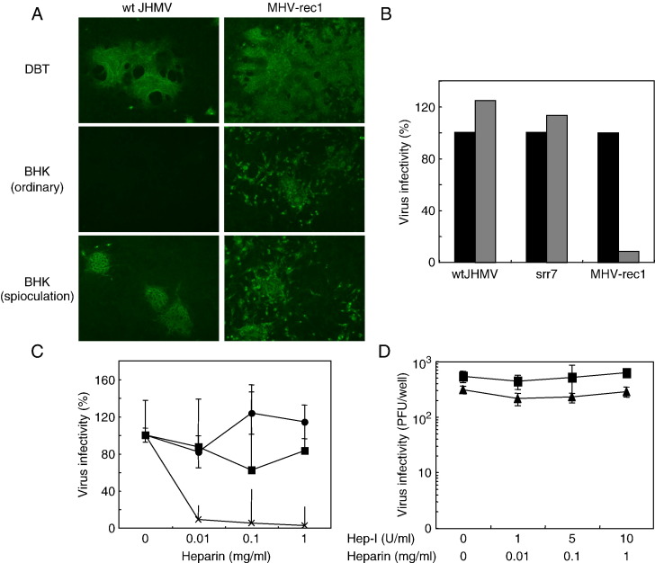
A highly neurovirulent mouse hepatitis virus (MHV) JHMV strain (wt) with receptor (MHVR)-independent infection activity and its low-virulent mutant srr7 without such activity were found to attach to MHVR-negative, non-permissive BHK cells. To identify the molecule that interacts with JHMV, we focused on heparan sulfate (HS) since it works as a receptor of a mutant MHV-rec1 that infects in an MHVR-independent fashion. The present study indicates that HS interacts with both wt JHMV and srr7 but it does not function as an entry receptor as it apparently does for MHV-rec1. Furthermore, HS failed to serve as an entry receptor in the MHVR-independent infection of wt JHMV, indicating that HS is not a host factor that wt JHMV utilizes in an MHVR-independent infection.


Figures

Fig. 1
Fig. 1
HS-binding sites on the S protein of wt JHMV and MHV-rec1. S proteins of wt JHMV and MHV-rec1 consisting of 1376 and 1331 amino acids, respectively, are depicted as boxes. The receptor binding site and transmembrane region are shown as closed and open boxes, respectively. Vertical lines in the boxes represent approximate locations of putative HS-binding motifs. Putative HS-binding consensus sequences (XBXBBX or XBXXBBBX; X = any amino acid, B = basic amino acid) are underlined. The S1/S2 cleavage site of wt JHMV is shown by a slash (/).
Fig. 2
Fig. 2
Binding of JHMV to BHK and BHK-R1 cells. (A) 105 PFU (ca. 107 genome copies) of wt JHMV and srr7 were inoculated onto BHK-R1 (black column) and BHK (gray column) cells and incubated at 4 °C for 1 h. After 3 washes, total RNA was recovered from cells and the copy number of attached virus was estimated by real-time PCR. (B) BHK cells infected with serially diluted 105 PFU of wt JHMV or srr7 as described in (A) were further incubated for 14 h in the presence (black column) or absence (gray column) of 50 nM soMHVR. The number of plaque was counted after staining with crystal violet. Error bars represent standard deviations of the results of three independent experiments.
Fig. 3
Fig. 3
Effects of heparinase (A–C) and heparin (D) treatment on virus attachment to BHK and BHK-R1 cells. (A) Effect of heparinase treatment on the amount of cell surface HS (a) and MHVR (b). Cells were treated with heparinase I (5 U/ml) or III (1 U/ml) at 37 °C for 1 h, and treated cells were stained with anti-HS or anti-MHVR MAbs followed by fluorescence-conjugated secondary antibodies. Fluorescence intensities were analyzed by FACSCalibur. (B) BHK cells were treated with either heparinase I (10 U/ml) or III (2 U/ml) at 37 °C for 1 h. Those cells were then inoculated with ca. 107 copies of JHMVs and incubated at 4 °C for 1 h. The copy number of cell-attached virus was estimated by real-time PCR using total RNA extracted from the infected cells. Values are represented as relative % against the copy number of viruses attached to cells that were not treated with heparinase. Error bars represent standard deviations of the results of three independent experiments. (C) Effect of heparinase treatment on srr7 attachment as examined by soMHVR-mediated infection. BHK and BHK-R1 cells were treated by heparinase I (Hep-1) (▴) or III (Hep-III) (●) at various concentrations (U/ml) at 37 °C for 1 h and were inoculated with 2 × 104 and 200 PFU of srr7, respectively. Cells were further incubated for 15 h in DMEM containing 1% FBS in the presence (BHK) or the absence (BHK-R1) of 50 nM soMHVR. The number of plaque was counted after staining with crystal violet. Error bars represent standard deviations of the results of three independent experiments. (D) Competition of the srr7 attachment by heparin. 104 or 200 PFU of srr7 was mixed with heparin and incubated at 4 °C for 1 h and mixtures were inoculated onto BHK (▪) and BHK-R1 (▴) cells, respectively. Cells were incubated for an additional 14 h at 37 °C in DMEM containing 1% FBS in the presence (BHK) or absence (BHK-R1) of 50 nM soMHVR. The number of plaque was counted after staining with crystal violet. Error bars represent standard deviations of the results of three independent experiments.
Fig. 4
Fig. 4
HS-mediated infection by JHMV or MHV-rec1. (A) Comparative infection of wt JHMV and MHV-rec1 onto MHVR-positive DBT or MHVR-negative BHK cells. 100 and 1000 PFU of each virus were infected onto DBT (DBT) and BHK cells, respectively, with an ordinary infection method; 37 °C for 1 h adsorption (BHK ordinary). 1000 PFU of each virus was spinoculated onto BHK cells (BHK spinoculation). Those infected cells were further incubated at 37 °C for 15 h. Infection was evaluated by immunofluorescence using the mixture of anti-JHMV-S MAbs followed by anti-mouse antibodies conjugated with FITC. (B) DBT cells were treated (gray column) or untreated (black column) with 5 U/ml of heparinase I, inoculated with 100 PFU of wt JHMV, srr7 or MHV-rec1 and incubated at 4 °C for 1 h. After 15 h of incubation at 37 °C, cells were stained with crystal violet. Virus titers are represented as the relative percentage of the virus titer obtained after infection of untreated cells. The result is representative of three independent experiments. (C) Effect of heparin on the infection of JHMV and MHV-rec1 on DBT cells. Two hundred plaque-forming units of wt JHMV (▪), srr7 (●) or MHV-rec1 (X) in 50 μl was mixed with an equal volume of heparin at a different concentration and the mixture was incubated at 4 °C for 1 h. The mixture was inoculated onto DBT cells, and cells were incubated at 4 °C for 1 h for virus adsorption. Cells were further incubated at 37 °C for 15 h and the number of plaque was counted after staining with crystal violet. Infectivity after treatment with heparin was shown as a percentage of the virus titer without heparin treatment. Error bars represent standard deviations of the results of three independent experiments. (D) Irrelevance of HS on MHVR-independent infection of wt JHMV. BHK cells were treated with various concentrations of heparinase I (Hep-1, U/ml) as described in the legend to Fig. 3C and then spinoculated with 104 PFU of wt JHMV. After 15 h of incubation at 37 °C, the number of plaque was counted (▴). 104 PFU of wt JHMV was mixed with various concentrations of heparin (mg/ml), as described in the legend to Fig. 3D, and the mixture was spinoculated onto BHK cells. The number of plaque was counted at 15 h after infection (▪). Error bars represent standard deviations of the results of three independent experiments.

Similar articles

Cited by

References

    1. Baric R.S., Yount B., Hensley L., Peel S.A., Chen W. Episodic evolution mediates interspecies transfer of a murine coronavirus. J. Virol. 1997;71(3):1946–1955. - PMC - PubMed
    1. Baric R.S., Sullivan E., Hensley L., Yount B., Chen W. Persistent infection promotes cross-species transmissibility of mouse hepatitis virus. J. Virol. 1999;73(1):638–649. - PMC - PubMed
    1. de Haan C.A., Li Z., te Lintelo E., Bosch B.J., Haijema B.J., Rottier P.J. Murine coronavirus with an extended host range uses heparan sulfate as an entry receptor. J. Virol. 2005;79(22):14451–14456. - PMC - PubMed
    1. de Haan C.A., Li Z., te Lintelo E., Li Z., Raaben M., Wurdinger T., Bosch B.J., Rottier P.J. Cooperative involvement of the S1 and S2 subunits of the murine coronavirus spike protein in receptor binding and extended host range. J. Virol. 2006;80(22):10909–10918. - PMC - PubMed
    1. de Parseval A., Elder J.H. Binding of recombinant feline immunodeficiency virus surface glycoprotein to feline cells: role of CXCR4, cell-surface heparans, and an unidentified non-CXCR4 receptor. J. Virol. 2001;75(10):4528–4539. - PMC - PubMed

Publication types