

Comparison of diagnostic accuracy of Heidelberg Retina Tomograph II and Heidelberg Retina Tomograph 3 to discriminate glaucomatous and nonglaucomatous eyes
- PMID: 17693382
- PMCID: PMC3928044
- DOI: 10.1016/j.ajo.2007.06.021
Comparison of diagnostic accuracy of Heidelberg Retina Tomograph II and Heidelberg Retina Tomograph 3 to discriminate glaucomatous and nonglaucomatous eyes
Abstract
Purpose: To compare the diagnostic accuracy of the Moorfields regression analysis (MRA), parameters, and glaucoma probability score (GPS) from Heidelberg Retinal Tomograph (HRT) 3 (Heidelberg Engineering, Heidelberg, Germany) with MRA and parameters from HRT II in discriminating glaucomatous and healthy eyes in subjects of African and European ancestry.
Design: Case-control institutional setting.
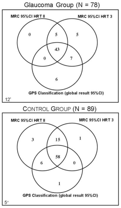
Methods: Seventy-eight glaucoma patients (44 of African ancestry, 34 of European ancestry) and 89 age-matched controls (46 of African ancestry, 33 European ancestry), defined by visual fields and self-reported race were included. Imaging was obtained with HRT II, and data were exported to a computer with the HRT 3 software using the same contour line. Area under the receiver operating characteristic (ROC) curves (AUCs), sensitivity, and specificity were evaluated for the entire group, the African ancestry group, and the European ancestry group separately. Mean disk area was compared between correctly and incorrectly diagnosed eyes by each technique.
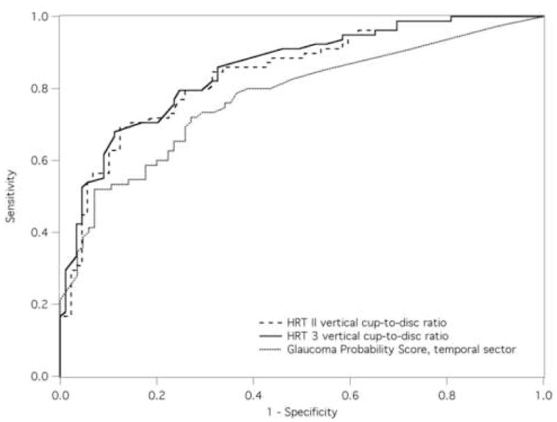
Results: Disk, cup, and rim areas from HRT 3 were lower than HRT II (P < .0001). AUC (sensitivity at 95% specificity) was 0.85 (54%) for vertical cup-to-disk ratio (VCDR) HRT 3, 0.84 (45%) for VCDR HRT II, and 0.81 (44%) for GPS at the temporal sector. MRA HRT 3 showed greater sensitivity but lower specificity than HRT II for the entire group, the African ancestry group, and the European ancestry group. GPS classification had the lowest specificity. Glaucomatous eyes incorrectly classified by GPS had smaller mean disk area (P = .0002); control eyes incorrectly classified had greater mean disk area (P = .015).
Conclusions: VCDR from HRT 3 showed higher sensitivity than HRT II and GPS for the entire group and for those of African ancestry and of European ancestry separately. Sensitivity of MRA improved in HRT 3 with some trade-off in specificity compared with MRA of HRT II. GPS yielded erroneous classification associated to optic disk size.
Figures

References
-
- Caprioli J, Park HJ, Ugurlu S, Hoffman D. Slope of the peripapillary nerve fiber layer surface in glaucoma. Invest Ophthalmol Vis Sci. 1998;39:2321–8. - PubMed
-
- Miglior S, Casula M, Guareschi M, Marchetti I, Iester M, Orzalesi N. Clinical ability of Heidelberg retinal tomograph examination to detect glaucomatous visual field changes. Ophthalmology. 2001;108:1621–7. - PubMed
-
- Wollstein G, Garway-Heath DF, Hitchings RA. Identification of early glaucoma cases with the scanning laser ophthalmoscope. Ophthalmology. 1998;105:1557–63. - PubMed
-
- Wollstein G, Garway-Heath DF, Fontana L, Hitchings RA. Identifying early glaucomatous changes. Comparison between expert clinical assessment of optic disc photographs and confocal scanning ophthalmoscopy. Ophthalmology. 2000;107:2272–7. - PubMed
-
- Zangwill LM, Bowd C, Berry CC, et al. Discriminating between normal and glaucomatous eyes using the Heidelberg Retina Tomograph, GDx Nerve Fiber Analyzer, and Optical Coherence Tomograph. Arch Ophthalmol. 2001;119:985–93. - PubMed
Publication types
MeSH terms
Grants and funding
LinkOut - more resources
Full Text Sources
Medical
