. 2007 Oct 19;282(42):30673-9.
doi: 10.1074/jbc.M703622200. Epub 2007 Aug 10.

Down syndrome critical region-1 is a transcriptional target of nuclear factor of activated T cells-c1 within the endocardium during heart development

Affiliations

Down syndrome critical region-1 is a transcriptional target of nuclear factor of activated T cells-c1 within the endocardium during heart development

Hai Wu et al. J Biol Chem. .

Abstract

Patients with Down syndrome have characteristic heart valve lesions resulting from endocardial cushion defects. The Down syndrome critical region 1 (DSCR1) gene, identified at the conserved trisomic 21 region in those patients, encodes a calcineurin inhibitor that inactivates nuclear factor of activated T cells (NFATc) activity. Here, we identify a regulatory sequence in the promoter region of human DSCR1 that dictates specific expression of a reporter gene in the endocardium, defined by the temporal and spatial expression of Nfatc1 during heart valve development. Activation of this evolutionally conserved DSCR1 regulatory sequence requires calcineurin and NFATc1 signaling in the endocardium. NFATc1 proteins bind to the regulatory sequence and trigger its enhancer activity. NFATc1 is sufficient to induce the expression of Dscr1 in cells that normally have undetectable or minimal NFATc1 or DSCR1. Pharmacologic inhibition of calcineurin or genetic Nfatc1 null mutation in mice abolishes the endocardial activity of this DSCR1 enhancer. Furthermore, in mice lacking endocardial NFATc1, the endogenous Dscr1 expression is specifically inhibited in the endocardium but not in the myocardium. Thus, our studies indicate that the DSCR1 gene is a direct transcriptional target of NFATc1 proteins within the endocardium during a critical window of heart valve formation.


Figures
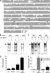
FIGURE 1
FIGURE 1. NFATc proteins bind and transactivate a conserved enhancer derived from the DSCR1 gene
A, alignment of part of the enhancer from human, mouse, and rat. The nucleotide immediately upstream of the start codon ATG of exon 4 is defined as position −1. Oligonucleotides (NREs) used in the gel shift assay are boxed. B, binding of NFATc proteins to the enhancer. [γ32P] ATP-labeled NRE oligonucleotides were incubated with nuclear extracts from mouse embryonic hearts. DNA-protein complex (arrow) was resolved by gel electrophoresis. For the NFAT, DNA complexes were disrupted after incubating nuclear protein extracts with anti-NFATc1 antibody. C, binding of NFATc proteins to intact and mutant NREs. The consensus NFAT binding sites (GGAAA) were substituted with CTAGC in the mutant NREs. For the formation of NFAT, DNA complexes (arrow) were disrupted for mutant NRE2 and −3. D, transactivation of the DSCR1 enhancer by calcineurin and NFATc1. CnA* and NFATc1 activated the enhancer in MEFs. E, NRE3 functions as an NFAT-dependent enhancer. Nine copies of intact or mutant NRE3 were cloned upstream of luciferase reporter. Transactivation of the mutant NRE3 (pGL3-Mut(NRE3)9) by calcineurin and NFATc1 is greatly reduced compared with intact NRE3 (pGL3-wt(NRE3)9).
FIGURE 2
FIGURE 2. The cDNA microarray analysis shows that NFATc1 activates endogenous DSCR1 expression
A, immunofluorescent staining shows nuclear localization NFATc1 proteins (red) in cells transfected with the constitutively nuclear NFATc1. B, Western blot analysis shows the expression of constitutively activated NFATc1 in REN cells after infection with adeno-NFATc1-IRES-GFP virus. REN cells infected with control GFP-only virus do not express detectable endogenous NFATc1 even with 10-fold protein loading. C, a graph showing that the ectopic expression of NFATc1 in REN cells markedly up-regulates DSCR1 mRNA expression (up to 11-fold, n = 4, p < 0.05). D, semiquantitative RT-PCR showing up-regulation of DSCR1 mRNA level in virally infected REN cells. Approximately 4-fold induction of DSCR1 mRNA by NFATc1 was observed.
FIGURE 3
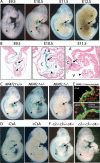
FIGURE 3. The expression of DSCR1-lacZ transgene in the endocardium requires calcineurin and NFATc1
A, transgenic embryos harboring the lacZ reporter driven by the DSCR1 enhancer. Images of whole-mount 5-bromo-4-chloro-3-indolyl-β-d-galactopyranoside (X-gal) staining of embryos at different developmental stages (E9.5 to E12.5) are shown. lacZ is expressed in both the heart (arrows) and neural tube (arrowheads). B, sections of DSCR1-lacZ embryonic hearts at E9.5, E10.5, and E11.5 are counterstained with nuclear fast red. Note that the lacZ is specifically expressed in endocardium of the cushion (arrows) and ventricle (arrowheads) at E9.5 and E10.5. Its expression is limited to the cushion endocardium at E11.5. A, atrium; C, cushion; V, ventricle. C, images of whole-mount X-gal staining of embryos at E10.5. Breeding of lacZ transgene into Nfatc1 null mutant background removes lacZ expression in the heart (arrows) but not in the neural tube (arrowheads). Nfatc1 heterozygous mutant retains lacZ expression. D, images of whole-mount X-gal staining of embryos at E9.5. Staged pregnant transgenic mice (E8) were subcutaneously injected with cyclosporin A (CsA) at 50 mg/kg body weight. Embryos were harvested and analyzed 36 h later. CsA treatment prevents the transgene expression in the heart (arrows) and neural tube (arrowheads). E, CsA-treated embryos showed cytoplasmic translocation of NFATc1 proteins in the cushion endocardium. Fluorescent immunostaining of NFATc1 proteins by anti-NFATc1 antibody (green) and nuclei by 7-amino-actinomycin D (red)is shown. F, NFATc2, c3, or c4 is not required to maintain DSCR1-lacZ expression in the endocardium (arrows). Breeding of lacZ transgene into Nfatc2/c3/c4 triple null mutant background removes lacZ expression in the neural tube (arrowheads) but not in the heart (arrows).
FIGURE 4
FIGURE 4. NFATc1 activates the expression of endogenous DSCR1 in the endocardium
A, the expression pattern of DSCR1 mRNA by in situ hybridization at E12.5. Dscr1 mRNA is expressed in both the myocardium (arrowheads) and cushion endocardium (arrows). Black dots, Dscr1 mRNA; blue, hematoxylin counterstain; *, valve leaflet. B, the expression of DSCR1 in the cushion endocardium (arrow) is abolished in the absence of NFATc1. DSCR1 expression in the myocardium (arrowhead) is preserved. *, valve leaflet. C, higher magnification of panel A showing abundant Dscr1 mRNA (black dots) expression in the cushion endocardium (arrows). D, higher magnification of panel B, showing minimal Dscr1 mRNA expression in the cushion endocardium of Nfatc1−/− embryos (arrows). E and F, PECAM-1 immunostaining of cushion endocardium of wild-type (E) and Nfatc1−/− embryos (F) at E12.5. Green, PECAM-1 proteins; blue,4′,6-diamidino-2-phenylindole nuclear stain. G, a working model illustrating a negative feedback loop that controls NFATc1 activity and DSCR1 expression within the endocardium. DSCR1 inhibits calcineurin and NFATc1 signaling required for its own expression. NFATc1 proteins activate the expression of DSCR1, a calcineurin inhibitor, to regulate its nuclear localization and activity.

Similar articles

Cited by

References

    1. Roper RJ, Reeves RH. PLoS Genet. 2006;2:e50. - PMC - PubMed
    1. Morris JK, Wald NJ, Watt HC. Prenat. Diagn. 1999;19:142–145. - PubMed
    1. Spencer K. Prenat. Diagn. 2001;21:788–789. - PubMed
    1. Ferencz C, Neill CA, Boughman JA, Rubin JD, Brenner JI, Perry LW. J. Pediatr. 1989;114:79–86. - PubMed
    1. Epstein CJ. Am. J. Med. Genet. Suppl. 1990;7:31–37. - PubMed

Publication types

MeSH terms

LinkOut - more resources