. 2007;35(16):5370-8.
doi: 10.1093/nar/gkm580. Epub 2007 Aug 9.

Ligand-induced folding of the thiM TPP riboswitch investigated by a structure-based fluorescence spectroscopic approach

Affiliations

Ligand-induced folding of the thiM TPP riboswitch investigated by a structure-based fluorescence spectroscopic approach

Kathrin Lang et al. Nucleic Acids Res. 2007.

Abstract

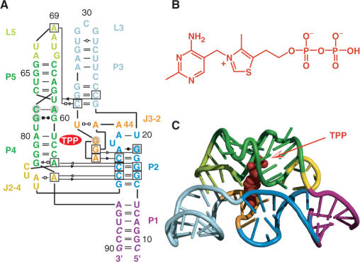
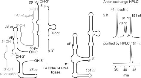
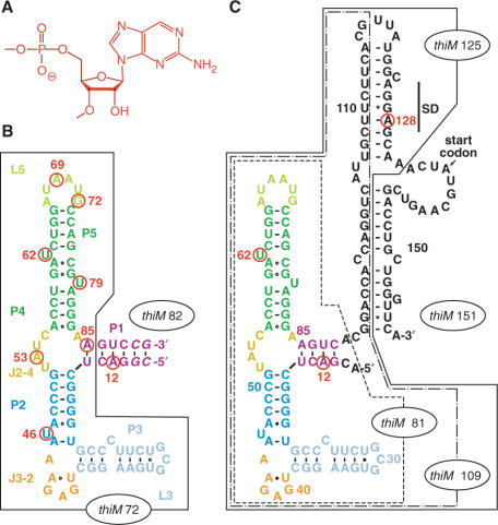
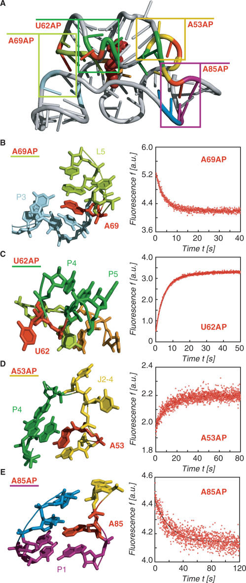
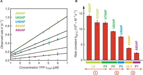
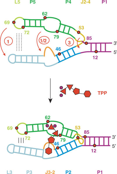
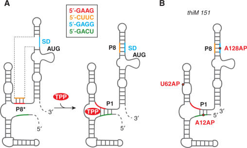
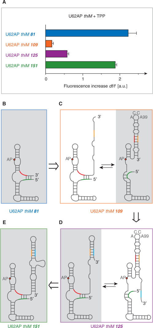
Riboswitches are genetic control elements within non-coding regions of mRNA. They consist of a metabolite-sensitive aptamer and an adjoining expression platform. Here, we describe ligand-induced folding of a thiamine pyrophosphate (TPP) responsive riboswitch from Escherichia coli thiM mRNA, using chemically labeled variants. Referring to a recent structure determination of the TPP/aptamer complex, each variant was synthesized with a single 2-aminopurine (AP) nucleobase replacement that was selected to monitor formation of tertiary interactions of a particular region during ligand binding in real time by fluorescence experiments. We have determined the rate constants for conformational adjustment of the individual AP sensors. From the 7-fold differentiation of these constants, it can be deduced that tertiary contacts between the two parallel helical domains (P2/J3-2/P3/L3 and P4/P5/L5) that grip the ligand's ends in two separate pockets, form significantly faster than the function-critical three-way junction with stem P1 fully developed. Based on these data, we characterize the process of ligand binding by an induced fit of the RNA and propose a folding model of the TPP riboswitch aptamer. For the full-length riboswitch domain and for shorter constructs that represent transcriptional intermediates, we have additionally evaluated ligand-induced folding via AP-modified variants and provide insights into the sequential folding pathway that involves a finely balanced equilibrium of secondary structures.


Figures

Figure 1.
Figure 1.
Structure of the thiM riboswitch aptamer from E. coli in complex with thiamine pyrophosphate (TPP) (14); (A) Secondary/tertiary structure presentation in Leontis–Westhof nomenclature (18); (B) Chemical formula of TPP; (C) Cartoon representation of the overall fold of the RNA/ligand complex.
Scheme 1.
Scheme 1.
Enzymatic ligation strategy used for preparation of the E. coli thiM riboswitch variants. The 151-nt construct was chemically synthesized in four pieces and ligated enzymatically.
Figure 2.
Figure 2.
ThiM TPP riboswitch sequences used in this study; individual nucleobase replacements by 2-aminopurine (AP) are indicated by a circle and the corresponding number of position in red; (A) Chemical formula of an AP nucleotide unit; (B) Aptamer sequence thiM 82 (the number in the term indicates the sequence length of 82 nt); the truncated aptamer version thiM 72 is boxed; the sequence refers to the one used for crystal structure determination by Serganov et al. (14) with a 6 bp instead of a 5 bp stem which was initially chosen for reasons of enzymatic ligation; (C) Aptamer-expression platform sequence thiM 151; the truncated versions thiM 81, thiM 109, thiM 125 are boxed and represent mimics of transcriptional intermediates. Note that thiM 81 variants with the completely natural 4-bp stem provide ligation and fluorescence behaviors comparable with thiM 82 variants (B).
Figure 3.
Figure 3.
Structural analysis of the thiM TPP/aptamer complex for replacements with single 2-aminopurines (AP) and fluorescence response of the corresponding riboswitch variants; (A) Cartoon presentation of the overall fold with boxes indicating the regions for close-ups in (B–E); (B) A69: local structural environment (left) and stopped-flow fluorescence response of the A69AP thiM 82 variant upon TPP addition (right); conditions: cRNA = 0.3 μM, cTPP = 1.5 μM, 50 mM KMOPS, 100 mM KCl, 2 mM MgCl2, 25°C, pH 7.5; mixing was performed with a stopped-flow apparatus; (C) Same for U62; (D) Same for A53 and (E) Same for A85.
Figure 4.
Figure 4.
Kinetic assessment of the conformational rearrangements of riboswitch AP-variants upon TPP binding based on fluorescence measurements; (A) Plot of observed rate k′ versus ligand concentration for five different thiM 82 AP variants as indicated. Observed rates were determined under pseudo-first-order conditions from at least three independent stopped-flow measurements. The slope of the plot yields the rate constant k; (B) Graph visualizing the different magnitudes of rate constants k for individual AP variants, and their correlation to the label position and the corresponding secondary structure element; circled numbers 1–3 provide a rough classification into fast, middle and slow rate constants.
Figure 5.
Figure 5.
Proposed model for ligand-induced folding of the thiM TPP riboswitch aptamer based on the 7-fold differentiation of rate constants observed for the individual AP variants. Upon TPP binding, base–base and base–backbone interactions between nucleotides of L5 and P3 are formed (circle 1). Recognition of the pyrophosphate moiety of TPP by the 3′-helical domain (P4/P5) occurs almost simultaneously with recognition of the pyrimidine moiety by the 5′-helical domain (J3-2) (circle 1 and circle 2). Formation of the three-way junction and closure of stem P1 result from this initial recognition/folding process and require significantly more time to be fully accomplished (circle 3). TPP is schematically drawn in red (phosphates, triangles; thiazole, pentagon; pyrimidine, hexagon; Mg2+ ions are shown as circles in magenta).
Figure 6.
Figure 6.
ThiM TPP riboswitch; (A) Original model of the in vivo response mechanism proposed by Winkler et al. (6); (B) Schematic presentation of the three thiM 151 AP variants (U62AP, A12AP and A128AP) used in the present study.
Figure 7.
Figure 7.
ThiM riboswitch and mimics of transcriptional intermediates with U62AP replacements; (A) Relative fluorescence increase of U62AP variants of 81, 109, 125 and 151 nt length. Conditions: cRNA = 0.3 μM, cTPP = 3 μM, 50 mM KMOPS, 100 mM KCl, 2 mM MgCl2, pH 7.5 at 25°C. Due to the weak fluorescence increase of thiM 109 and thiM 125, TPP binding seems to be hindered, while for the full-length thiM 151 variant, ligand-binding capacity is well comparable with the aptamer domain alone; a likely explanation are alternative competing secondary structures that are binding incompetent: (BE) and Supplementary Figure S10.

Similar articles

Cited by

References

    1. Mandal M, Breaker RR. Gene regulation by riboswitches. Nat. Rev. Mol. Cell Biol. 2004;5:451–463. - PubMed
    1. Winkler WC, Breaker RR. Genetic control by metabolite-binding riboswitches. Chembiochem. 2003;4:1024–1032. - PubMed
    1. Soukup JK, Soukup GA. Riboswitches exert genetic control through metabolite-induced conformational change. Curr. Opin. Struct. Biol. 2004;14:344–349. - PubMed
    1. Sashital DG, Butcher SE. Flipping off the riboswitch: RNA structures that control gene expression. ACS Chem. Biol. 2006;1:341–345. - PubMed
    1. Nudler E, Mironov AS. The riboswitch control of bacterial metabolism. Trends Biochem. Sci. 2004;29:11–17. - PubMed

Publication types