. 2007 Nov 15;93(10):3421-33.
doi: 10.1529/biophysj.107.110031. Epub 2007 Aug 10.

Modeling hypertrophic IP3 transients in the cardiac myocyte

Affiliations

Modeling hypertrophic IP3 transients in the cardiac myocyte

Michael Cooling et al. Biophys J. .

Abstract

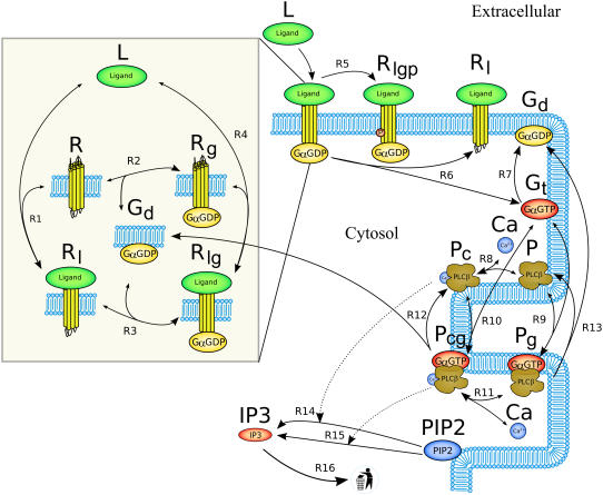
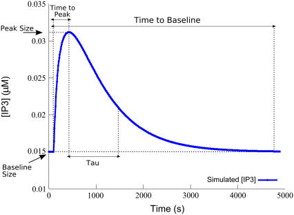
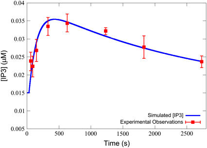
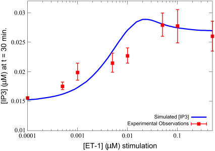
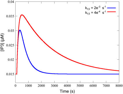
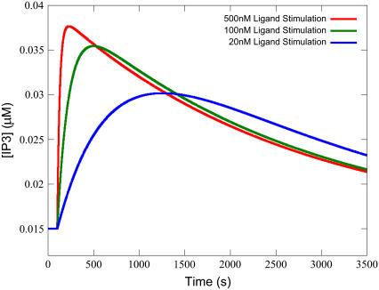
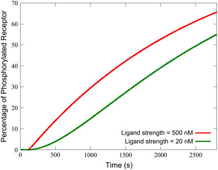
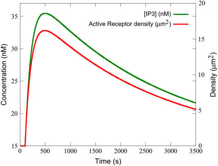
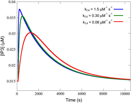
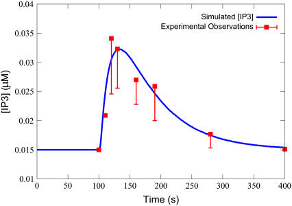
Cardiac hypertrophy is a known risk factor for heart disease, and at the cellular level is caused by a complex interaction of signal transduction pathways. The IP3-calcineurin pathway plays an important role in stimulating the transcription factor NFAT which binds to DNA cooperatively with other hypertrophic transcription factors. Using available kinetic data, we construct a mathematical model of the IP3 signal production system after stimulation by a hypertrophic alpha-adrenergic agonist (endothelin-1) in the mouse atrial cardiac myocyte. We use a global sensitivity analysis to identify key controlling parameters with respect to the resultant IP3 transient, including the phosphorylation of cell-membrane receptors, the ligand strength and binding kinetics to precoupled (with G(alpha)GDP) receptor, and the kinetics associated with precoupling the receptors. We show that the kinetics associated with the receptor system contribute to the behavior of the system to a great extent, with precoupled receptors driving the response to extracellular ligand. Finally, by reparameterizing for a second hypertrophic alpha-adrenergic agonist, angiotensin-II, we show that differences in key receptor kinetic and membrane density parameters are sufficient to explain different observed IP3 transients in essentially the same pathway.


Figures

FIGURE 1
FIGURE 1
Reaction scheme of the IP3 production system. The extracellular ligand (L) binds to receptors (R), whether precoupled with GαGDP (Gd, yielding Rlg) or not (Rl). Fully activated receptors (Rlg) release GαGTP (Gt), which, along with calcium (Ca), stimulates PLCβ (P). In the unstimulated state, PLCβ-Ca2+ (Pc) hydrolyzes PIP2 to produce IP3 via reaction R14. When stimulated, PLCβ-Ca2+-GαGTP (Pcg) hydrolyses PIP2 at a faster rate via reaction R15. Free IP3 is degraded via reaction R16.
FIGURE 2
FIGURE 2
IP3 transient-based objective measures. Graphical depiction of the measures used to define the objective functions for the sensitivity analysis. Tau is the time point at which the transient dips beneath 1/e × amplitude.
FIGURE 3
FIGURE 3
IP3 transient curve on ET-1 stimulation. The simulated IP3 transient closely matches experimental observations (40) on application of 100 nM ET-1. Simulations were performed with the equations as in Table 3 and the parameters and initial conditions as in Table 4. Additionally, ts = 30 s, and Ls = 0.100 μM.
FIGURE 4
FIGURE 4
IP3 dose-response curve on ET-1 stimulation. The model also closely matches experimental observations (40) for the concentration of IP3, after 30 min, on stimulation by various concentrations of ET-1. Simulations were performed with the equations as in Table 3 and the parameters and initial conditions as in Table 4. Additionally, ts = 100 s, and the model was run until (tts) = 1800 s (30 min of stimulation) for each of Ls = 1 × 10⁻⁴, 5 × 10⁻⁴, 1 × 10⁻³, 5 × 10⁻³, 1 × 10⁻², 5 × 10², 1 × 10⁻¹, and 5 × 10⁻¹ μM.
FIGURE 5
FIGURE 5
Decreased kf,5 results in a higher peak and a longer time-to-baseline from peak. Simulations were performed with the equations as in Table 3 and the parameters and initial conditions as in Table 4. Additionally, ts = 100 s, Ls = 0.100 μM, and kf,5 was varied as depicted in the legend.
FIGURE 6
FIGURE 6
Decreasing ligand strength strongly effects Time-to-Peak. Lower ligand strength produces a slower rate of active receptor production, thus the transient takes more time to achieve its maximum. Simulations were performed with the equations as in Table 3 and the parameters and initial conditions as in Table 4. Additionally, ts = 100 s and Ls was varied as depicted in the legend.
FIGURE 7
FIGURE 7
Higher ligand strength more readily increases phosphorylated receptor density. Although the long-term gradients for phosphorylated receptor density are similar for both high and low concentrations of ligand strength, a higher strength yields a much larger initial gain. Simulations were performed with the equations as in Table 3 and the parameters and initial conditions as in Table 4. Additionally, ts = 100 s and Ls was varied as depicted in the legend.
FIGURE 8
FIGURE 8
The IP3 transient follows the active receptor transient. Simulations were performed with the equations as in Table 3 and the parameters and initial conditions as in Table 4. Additionally, ts = 100 s and Ls = 0.100 μM.
FIGURE 9
FIGURE 9
Higher kf,4 reduces the Tau-to-Tail Ratio. Increased kf,4 gives sharper peaks, but longer tails from the peak, and a lower Tau-to-Tail Ratio compared to flatter transients with a higher ratio when kf,4 is decreased. Simulations were performed with the equations as in Table 3 and the parameters and initial conditions as in Table 4. Additionally, ts = 100 s, Ls = 0.100 μM, and kf,4 was varied as depicted in the legend.
FIGURE 10
FIGURE 10
Simulated fit to the observations of Abdellatif et al. (39). The simulated transient matches the observations (39) for stimulation with 100 nM of Ang-2. Simulations were performed with the equations as in Table 3 and the parameters and initial conditions as in Table 4, and the Ang-2 values listed in Table 2. Additionally, Ls = 100 μM and ts = 100 s.

Similar articles

Cited by

References

    1. Heineke, J., and J. D. Molkentin. 2006. Regulation of cardiac hypertrophy by intracellular signaling pathways. Nat. Rev. Mol. Cell Biol. 7:589–600. - PubMed
    1. Molkentin, J. D., J.-R. Lu, C. L. Antos, B. Markham, J. Richardson, J. Robbins, S. R. Grant, and E. N. Olson. 1998. A calcineurin-dependent transcriptional pathway for cardiac hypertrophy. Cell. 93:215–228. - PMC - PubMed
    1. Sugden, P. H. 1999. Signaling in myocardial hypertrophy: life after calcineurin? Circ. Res. 84:633–646. - PubMed
    1. Bare, D. J., C. S. Kettlun, M. Liang, D. M. Bers, and G. A. Mignery. 2005. Cardiac type 2 inositol 1,4,5-trisphosphate receptor: interaction and modulation by calcium/calmodulin-dependent protein kinase II. J. Biol. Chem. 280:15912–15920. - PubMed
    1. Hardt, S. E., and J. Sadoshima. 2002. Glycogen synthase kinase-3β: a novel regulator of cardiac hypertrophy and development. Circ. Res. 90:1055–1063. - PubMed

LinkOut - more resources