. 2007 Oct;189(20):7367-75.
doi: 10.1128/JB.00590-07. Epub 2007 Aug 10.

Characterization of riboflavin (vitamin B2) transport proteins from Bacillus subtilis and Corynebacterium glutamicum

Affiliations

Characterization of riboflavin (vitamin B2) transport proteins from Bacillus subtilis and Corynebacterium glutamicum

Christian Vogl et al. J Bacteriol. 2007 Oct.

Abstract

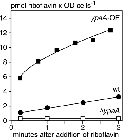
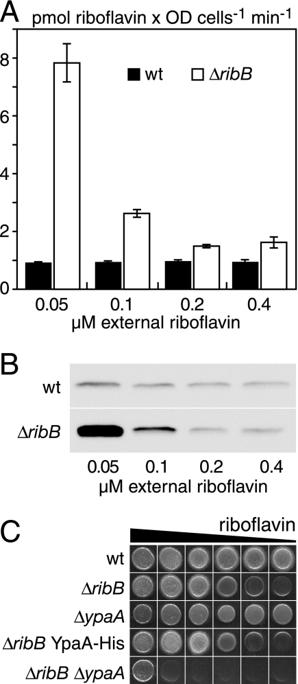
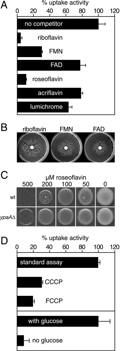
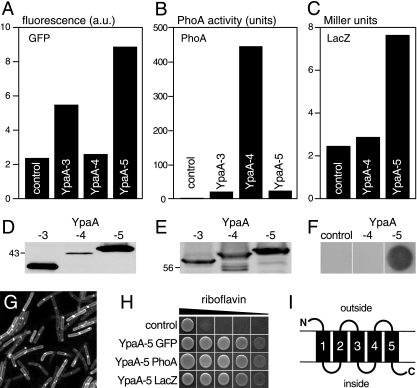
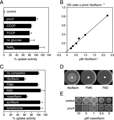
Riboflavin (vitamin B(2)) is the direct precursor of the flavin cofactors flavin mononucleotide and flavin adenine dinucleotide, essential components of cellular biochemistry. In this work we investigated the unrelated proteins YpaA from Bacillus subtilis and PnuX from Corynebacterium glutamicum for a role in riboflavin uptake. Based on the regulation of the corresponding genes by a riboswitch mechanism, both proteins have been predicted to be involved in flavin metabolism. Moreover, their primary structures suggested that these proteins integrate into the cytoplasmic membrane. We provide experimental evidence that YpaA is a plasma membrane protein with five transmembrane domains and a cytoplasmic C terminus. In B. subtilis, riboflavin uptake was increased when ypaA was overexpressed and abolished when ypaA was deleted. Riboflavin uptake activity and the abundance of the YpaA protein were also increased when riboflavin auxotrophic mutants were grown in limiting amounts of riboflavin. YpaA-mediated riboflavin uptake was sensitive to protonophors and reduced in the absence of glucose, demonstrating that the protein requires metabolic energy for substrate translocation. In addition, we demonstrate that PnuX from C. glutamicum also is a riboflavin transporter. Transport by PnuX was not energy dependent and had high apparent affinity for riboflavin (K(m) 11 microM). Roseoflavin, a toxic riboflavin analog, appears to be a substrate of PnuX and YpaA. We propose to designate the gene names ribU for ypaA and ribM for pnuX to reflect that the encoded proteins function in riboflavin uptake and that the genes have different phylogenetic origins.


Figures

FIG. 1.
FIG. 1.
Riboflavin uptake in B. subtilis depends on YpaA. Uptake assays were performed with B. subtilis strains grown in riboflavin-free CSEG medium. The strains were either wild type (wt), contained a plasmid for overexpression of ypaA (ypaA-OE), or had a deletion of the ypaA gene (ΔypaA::Kanr). The cells were resuspended in transport buffer (50 mM K2HPO4/KH2PO4, 50 mM MgCl2, pH 7.0), energized with 1 mM glucose, and the experiment was started by adding [14C]riboflavin for a final concentration of 1.6 μM. Aliquots were withdrawn, filtered, and extensively washed with water, and the radioactivity was determined by liquid scintillation counting. The experiment was performed twice with similar results.
FIG. 2.
FIG. 2.
Regulation of riboflavin uptake activity by external riboflavin. (A) B. subtilis wild-type cells or a B. subtilis ΔribB:Ermr mutant cells were grown in synthetic CSEG medium to which riboflavin was added to give the desired concentrations. Standard uptake experiments with [14C]riboflavin were performed, and the uptake velocities were calculated for the first 4 minutes of the experiments. Bars represent means, and error bars indicate standard deviation values of three independent experiments. (B) YpaA was genomically tagged with a C-terminal his tag either in a wild-type strain or a strain carrying the ΔribB:Tetr disruption. After growth in CSEG medium containing the indicated concentrations of riboflavin, the cells were collected and membrane extracts were separated by sodium dodecyl sulfate-polyacrylamide gel electrophoresis, followed by transfer to a nitrocellulose membrane and detection with the Penta-His mouse monoclonal antibody. (C) Growth assays on CSEG plates containing (from left to right) 20 mg/liter, 2 mg/liter, 200 μg/liter, 20 μg/liter, 2 μg/liter, or 200 ng/liter riboflavin were performed. Each spot contained approximately 50,000 cells, and the plates were incubated for 1 day at 37°C. The exact genotype of the strains was ΔribB:Ermr, ΔypaA:Kanr, and the double mutant carried both these deletions.
FIG. 3.
FIG. 3.
Characterization of the activity and energy requirements of YpaA. (A) Uptake of [14C]riboflavin (initial outside concentration, 1.6 μM) was studied in B. subtilis ΔribB:Ermr mutants grown in CSEG minimal medium containing 20 μg/liter riboflavin. Uptake was determined in the absence of competitors (mean uptake velocity, 8.29 pmol riboflavin × OD of cells−1 min−1; corresponds to 100%) or in the presence of a 10-fold excess (16 μM) of the given compounds. Bars represent means, and error bars indicate standard deviation (SD) values of three independent determinations. (B) B. subtilis ΔribB:Ermr mutants were plated on riboflavin-free CSEG minimal medium to produce a lawn. Next, a filter disc was placed in the middle of the plates and impregnated with 20 μl of a solution containing 540 μM riboflavin, FMN, or FAD. The area of growth around the disc was scored after incubation for 1 day at 37°C and had a diameter of 76 mm for riboflavin, 50 mm for FMN, and 42 mm for FAD. (C) B. subtilis wild-type cells (wt) or the ΔypaA::Kanr mutant were spotted on CSEG plates containing the indicated concentration of roseoflavin. Growth was recorded after incubation for 1 day at 42°C. (D) Uptake of [14C]riboflavin was determined using a ΔribB::Ermr mutant grown in CSEG medium containing 20 μg/liter riboflavin. The transport activity was determined without further additions (uptake velocity, 7.13 pmol riboflavin × OD cells−1 min−1; corresponds to 100%) or after adding CCCP (130 μM) or FCCP (130 μM) 3 min before starting the test by addition of the labeled substrate. To investigate if glucose stimulated riboflavin uptake, we stored the cells on ice for 2 hours in transport buffer. After this time, uptake activity was measured with [14C]riboflavin in the absence or presence of 1 mM glucose. In this experiment, 100% corresponds to an uptake activity of 5.1 pmol riboflavin × OD cells−1 min−1. Bars represent means, and error bars indicate SD values of three independent experiments.
FIG. 4.
FIG. 4.
Membrane topology of YpaA. The topology of YpaA was analyzed by generating a C-terminal fusion of YpaA to GFP (A), PhoA (B), or LacZ (C) in the vector pDG148-Stu. According to the prediction of the TMpred program (16), the fusion proteins contained either three (YpaA-3; fusion after amino acid 103) or four (YpaA-4; fusion after amino acid 146) transmembrane domains of YpaA or the full-length protein (YpaA-5). Plasmids were transformed into B. subtilis wild-type cells, which were grown in LB medium and induced with IPTG prior to the analysis. The activity of GFP was determined by fluorescence spectroscopy of intact cells. The fluorescence of vector control cells was used to normalize the measurements. Arbitrary units (a.u.) are given. The activity of alkaline phosphatase was assayed in intact cells with p-nitrophenyl phosphate using a standard protocol (27). The activity of β-galactosidase is presented as Miller units and was assayed with ortho-nitrophenyl-β-d-galactoside as a substrate (32). (D) The GFP-fusion proteins used in panel A were detected in a Western blot assay with anti-GFP serum. The calculated molecular masses of the GFP fusions were as follows: 38.2 kDa for YpaA-3, 42.7 kDa for YpaA-4, and 47.6 kDa for YpaA-5. (E) A similar analysis was performed for the cells used in panel B. The calculated molecular masses of the PhoA fusions were as follows: 59.1 kDa for YpaA-3, 63.7 kDa for YpaA-4, and 68.7 kDa for YpaA-5. (F) Cells expressing fusions of LacZ to YpaA-4 and YpaA-5 were plated on an SP plate containing phleomycin and 0.5 mM IPTG. After growth, they were covered with X-Gal containing agarose and left for 3 hours at 37°C. The cells with the YpaA-5 construct turned blue, whereas the other cells did not show a color reaction. (G) The cells expressing YpaA-5 GFP were also visualized using confocal microscopy. (H) We tested the functionality of the fusion constructs used in panels A to G in B. subtilis ΔribB::Ermr ΔypaA::Kanr mutants. Cells containing the indicated plasmids were spotted on IPTG-containing CSEG plates supplemented with (from left to right) 20 mg/liter, 2 mg/liter, 200 μg/liter, 20 μg/liter, or 2 μg/liter riboflavin. Growth was recorded 1 day after plating. Fusion proteins containing YpaA-3 or YpaA-4 did not complement the growth defect of the ΔribB mutant. (I) A model of the transmembrane topology of YpaA is shown.
FIG. 5.
FIG. 5.
Characterization of PnuX from Corynebacterium glutamicum as a riboflavin transporter. All uptake experiments were performed in the presence of 1 mM glucose with E. coli BL21(DE3) expressing a 3HA-tagged version of pnuX from plasmid pET21a(+) or harboring an empty pET21a(+) plasmid (control). (A) E. coli BL21 cells were either measured directly (standard assay; 100% corresponds to an activity of 5.57 pmol riboflavin × the OD in cells−1 min−1), in the presence of the protonophores CCCP (130 μM) or FCCP (130 μM), without glucose, or after the addition of 1 mM sodium azide. The glucose dependency was assayed as in Fig. 3D. (B) pET21a(+)-pnuX-3HA-transformed cells were subjected to uptake experiments in the presence of various concentrations of [14C]riboflavin. The graph represents the Lineweaver-Burk plot of an experiment from which a Km value of 11 μM was calculated. Two repetitions of this experiment yielded Km values of 5 μM and 17 μM, respectively. (C) Uptake experiments were performed with 2.2 μM [14C]riboflavin in the presence of a 10-fold excess of riboflavin or riboflavin analogs. (A and C), bars represent means and error bars indicate standard deviation values of three independent determinations. The uptake activity in the absence of competitors was 5.76 pmol riboflavin × OD cells−1 min−1 (100%). (D) E. coli ribB11 mutants were plated on riboflavin-free M9 minimal medium to produce a lawn and growth was assayed as in Fig. 3B. The area of growth had a diameter 53 mm for riboflavin and 18 mm for FMN. FAD produced no growth, even after incubation for one more day. (E) E. coli DH5α cells expressing pnuX from plasmid pDG148-pnuX-6His (pnuX) or containing an empty pDG148 plasmid (control) were spotted on M9 minimal medium plates containing the indicated concentrations of roseoflavin. Growth was recorded after 1 day at 37°C.

Similar articles

Cited by

References

    1. Bacher, A., S. Eberhardt, M. Fischer, K. Kis, and G. Richter. 2000. Biosynthesis of vitamin B2 (riboflavin). Annu. Rev. Nutr. 20:153-167. - PubMed
    1. Bandrin, S. V., P. M. Rabinovich, and A. I. Stepanov. 1993. Three linkage groups involved in riboflavin biosynthesis in E. coli. Sovi. Genet. 19:1103-1109. - PubMed
    1. Banerjee, R., and A. Batschauer. 2005. Plant blue-light receptors. Planta 220:498-502. - PubMed
    1. Burgess, C. M., D. J. Slotboom, E. R. Geertsma, R. H. Duurkens, B. Poolman, and D. van Sinderen. 2006. The riboflavin transporter RibU in Lactococcus lactis: molecular characterization of gene expression and the transport mechanism. J. Bacteriol. 188:2752-2760. - PMC - PubMed
    1. Cecchini, G., M. Perl, J. Lipsick, T. P. Singer, and E. B. Kearney. 1979. Transport and binding of riboflavin by Bacillus subtilis. J. Biol. Chem. 254:7295-7301. - PubMed

Publication types

MeSH terms

LinkOut - more resources