

Defining the stressome of Mycobacterium avium subsp. paratuberculosis in vitro and in naturally infected cows
- PMID: 17693514
- PMCID: PMC2168719
- DOI: 10.1128/JB.00780-07
Defining the stressome of Mycobacterium avium subsp. paratuberculosis in vitro and in naturally infected cows
Abstract
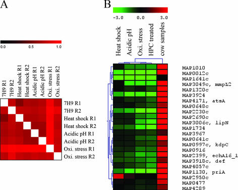
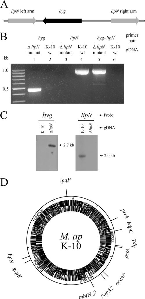
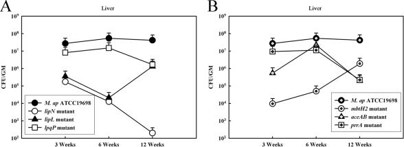
Mycobacterium avium subsp. paratuberculosis causes an enteric infection in cattle, with a great impact on the dairy industry in the United States and worldwide. Characterizing the gene expression profile of M. avium subsp. paratuberculosis exposed to different stress conditions, or shed in cow feces, could improve our understanding of the pathogenesis of M. avium subsp. paratuberculosis. In this report, the stress response of M. avium subsp. paratuberculosis on a genome-wide level (stressome) was defined for the first time using DNA microarrays. Expression data analysis revealed unique gene groups of M. avium subsp. paratuberculosis that were regulated under in vitro stressors while additional groups were regulated in the cow samples. Interestingly, acidic pH induced the regulation of a large number of genes (n=597), suggesting the high sensitivity of M. avium subsp. paratuberculosis to acidic environments. Generally, responses to heat shock, acidity, and oxidative stress were similar in M. avium subsp. paratuberculosis and Mycobacterium tuberculosis, suggesting common pathways for mycobacterial defense against stressors. Several sigma factors (e.g., sigH and sigE) were differentially coregulated with a large number of genes depending on the type of each stressor. Subsequently, we analyzed the virulence of six M. avium subsp. paratuberculosis mutants with inactivation of differentially regulated genes using a murine model of paratuberculosis. Both bacterial and histopathological examinations indicated the attenuation of all gene mutants, especially those selected based on their expression in the cow samples (e.g., lipN). Overall, the employed approach profiled mycobacterial genetic networks triggered by variable stressors and identified a novel set of putative virulence genes. A similar approach could be applied to analyze other intracellular pathogens.
Figures

Similar articles
-
Key role for the alternative sigma factor, SigH, in the intracellular life of Mycobacterium avium subsp. paratuberculosis during macrophage stress.Infect Immun. 2013 Jun;81(6):2242-57. doi: 10.1128/IAI.01273-12. Epub 2013 Apr 8. Infect Immun. 2013. PMID: 23569115 Free PMC article.
-
Characterization of genetic differences between Mycobacterium avium subsp. paratuberculosis type I and type II isolates.J Clin Microbiol. 2003 Nov;41(11):5215-23. doi: 10.1128/JCM.41.11.5215-5223.2003. J Clin Microbiol. 2003. PMID: 14605167 Free PMC article.
-
Virulence and immunity orchestrated by the global gene regulator sigL in Mycobacterium avium subsp. paratuberculosis.Infect Immun. 2014 Jul;82(7):3066-75. doi: 10.1128/IAI.00001-14. Epub 2014 May 5. Infect Immun. 2014. PMID: 24799632 Free PMC article.
-
Oligonucleotide microarray technology and its application to Mycobacterium avium subsp. paratuberculosis research: a review.Mol Biotechnol. 2009 May;42(1):30-40. doi: 10.1007/s12033-008-9137-5. Epub 2009 Jan 6. Mol Biotechnol. 2009. PMID: 19130317 Review.
-
SigE: A master regulator of Mycobacterium tuberculosis.Front Microbiol. 2023 Mar 7;14:1075143. doi: 10.3389/fmicb.2023.1075143. eCollection 2023. Front Microbiol. 2023. PMID: 36960291 Free PMC article. Review.
Cited by
-
Biomarkers for Early Stages of Johne's Disease Infection and Immunization in Goats.Front Microbiol. 2018 Sep 28;9:2284. doi: 10.3389/fmicb.2018.02284. eCollection 2018. Front Microbiol. 2018. PMID: 30323794 Free PMC article.
-
Mycobacterium avium subsp. Paratuberculosis and the expression of selected virulence and pathogenesis genes in response to 6°C, 65°c and ph 2.0.Braz J Microbiol. 2011 Apr;42(2):807-17. doi: 10.1590/S1517-838220110002000049. Epub 2011 Jun 1. Braz J Microbiol. 2011. PMID: 24031696 Free PMC article.
-
Lymphoproliferative and gamma interferon responses to stress-regulated Mycobacterium avium subsp. paratuberculosis recombinant proteins.Clin Vaccine Immunol. 2014 Jun;21(6):831-7. doi: 10.1128/CVI.00775-13. Epub 2014 Apr 2. Clin Vaccine Immunol. 2014. PMID: 24695774 Free PMC article.
-
mosR, a novel transcriptional regulator of hypoxia and virulence in Mycobacterium tuberculosis.J Bacteriol. 2009 Oct;191(19):5941-52. doi: 10.1128/JB.00778-09. Epub 2009 Jul 31. J Bacteriol. 2009. PMID: 19648248 Free PMC article.
-
Characterization of early immune responses elicited by live and inactivated vaccines against Johne's disease in goats.Front Vet Sci. 2023 Jan 9;9:1046704. doi: 10.3389/fvets.2022.1046704. eCollection 2022. Front Vet Sci. 2023. PMID: 36699320 Free PMC article.
References
-
- Ayele, W. Y., M. Macháèková, and I. Pavlík. 2001. The transmission and impact of paratuberculosis infection in domestic and wild ruminants. Vet. Med. 6-7:205-224.
-
- Bardarov, S., M. S. Pavelka, V. Sambandamurthy, M. Larsen, J. Tufariello, J. Chan, G. Hatfull, and W. R. Jacobs. 2002. Specialized transduction: an efficient method for generating marked and unmarked targeted gene disruptions in Mycobacterium tuberculosis, M. bovis BCG and M. smegmatis. Microbiology 148:3007-3017. - PubMed
-
- Betts, J. C., P. T. Lukey, L. C. Robb, R. A. McAdam, and K. Duncan. 2002. Evaluation of a nutrient starvation model of Mycobacterium tuberculosis persistence by gene and protein expression profiling. Mol. Microbiol. 43:717-731. - PubMed
Publication types
MeSH terms
Substances
LinkOut - more resources
Full Text Sources
Other Literature Sources
Molecular Biology Databases
