

Short-tailed stx phages exploit the conserved YaeT protein to disseminate Shiga toxin genes among enterobacteria
- PMID: 17693515
- PMCID: PMC2168440
- DOI: 10.1128/JB.00824-07
Short-tailed stx phages exploit the conserved YaeT protein to disseminate Shiga toxin genes among enterobacteria
Abstract
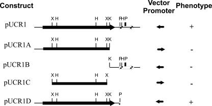
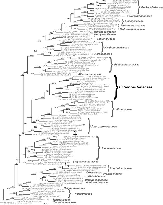
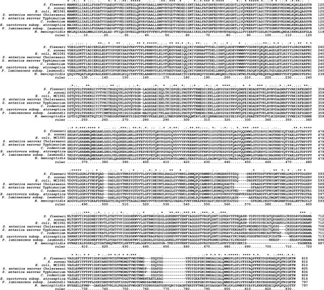
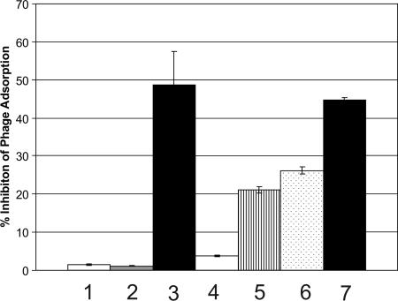
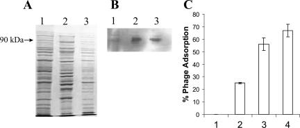
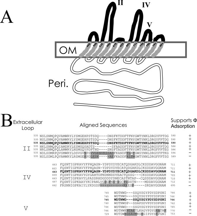
Infection of Escherichia coli by Shiga toxin-encoding bacteriophages (Stx phages) was the pivotal event in the evolution of the deadly Shiga toxin-encoding E. coli (STEC), of which serotype O157:H7 is the most notorious. The number of different bacterial species and strains reported to produce Shiga toxin is now more than 500, since the first reported STEC infection outbreak in 1982. Clearly, Stx phages are spreading rapidly, but the underlying mechanism for this dissemination has not been explained. Here we show that an essential and highly conserved gene product, YaeT, which has an essential role in the insertion of proteins in the gram-negative bacterial outer membrane, is the surface molecule recognized by the majority (ca. 70%) of Stx phages via conserved tail spike proteins associated with a short-tailed morphology. The yaeT gene was initially identified through complementation, and its role was confirmed in phage binding assays with and without anti-YaeT antiserum. Heterologous cloning of E. coli yaeT to enable Stx phage adsorption to Erwinia carotovora and the phage adsorption patterns of bacterial species possessing natural yaeT variants further supported this conclusion. The use of an essential and highly conserved protein by the majority of Stx phages is a strategy that has enabled and promoted the rapid spread of shigatoxigenic potential throughout multiple E. coli serogroups and related bacterial species. Infection of commensal bacteria in the mammalian gut has been shown to amplify Shiga toxin production in vivo, and the data from this study provide a platform for the development of a therapeutic strategy to limit this YaeT-mediated infection of the commensal flora.
Figures
Similar articles
-
Comparative genomics and stx phage characterization of LEE-negative Shiga toxin-producing Escherichia coli.Front Cell Infect Microbiol. 2012 Nov 7;2:133. doi: 10.3389/fcimb.2012.00133. eCollection 2012. Front Cell Infect Microbiol. 2012. PMID: 23162798 Free PMC article.
-
Diversity of stx2 converting bacteriophages induced from Shiga-toxin-producing Escherichia coli strains isolated from cattle.Microbiology (Reading). 2004 Sep;150(Pt 9):2959-2971. doi: 10.1099/mic.0.27188-0. Microbiology (Reading). 2004. PMID: 15347754
-
Comparison of Escherichia coli Isolates from humans, food, and farm and companion animals for presence of Shiga toxin-producing E. coli virulence markers.Foodborne Pathog Dis. 2004 Fall;1(3):178-84. doi: 10.1089/fpd.2004.1.178. Foodborne Pathog Dis. 2004. PMID: 15992278
-
Shiga toxins and stx phages: highly diverse entities.Microbiology (Reading). 2015 Mar;161(Pt 3):451-62. doi: 10.1099/mic.0.000003. Epub 2014 Dec 5. Microbiology (Reading). 2015. PMID: 25479836 Review.
-
A Toxic Environment: a Growing Understanding of How Microbial Communities Affect Escherichia coli O157:H7 Shiga Toxin Expression.Appl Environ Microbiol. 2020 Nov 24;86(24):e00509-20. doi: 10.1128/AEM.00509-20. Print 2020 Nov 24. Appl Environ Microbiol. 2020. PMID: 32358004 Free PMC article. Review.
Cited by
-
Shiga toxin-producing Escherichia coli O104:H4: a new challenge for microbiology.Appl Environ Microbiol. 2012 Jun;78(12):4065-73. doi: 10.1128/AEM.00217-12. Epub 2012 Apr 13. Appl Environ Microbiol. 2012. PMID: 22504816 Free PMC article. Review.
-
Comparative genomics of Shiga toxin encoding bacteriophages.BMC Genomics. 2012 Jul 16;13:311. doi: 10.1186/1471-2164-13-311. BMC Genomics. 2012. PMID: 22799768 Free PMC article.
-
Mobile effector proteins on phage genomes.Bacteriophage. 2012 Jul 1;2(3):139-148. doi: 10.4161/bact.21658. Bacteriophage. 2012. PMID: 23275865 Free PMC article.
-
CdiA Effectors from Uropathogenic Escherichia coli Use Heterotrimeric Osmoporins as Receptors to Recognize Target Bacteria.PLoS Pathog. 2016 Oct 10;12(10):e1005925. doi: 10.1371/journal.ppat.1005925. eCollection 2016 Oct. PLoS Pathog. 2016. PMID: 27723824 Free PMC article.
-
Isolation and identification of an Enterobacter cloacae strain producing a novel subtype of Shiga toxin type 1.J Clin Microbiol. 2014 Jul;52(7):2346-51. doi: 10.1128/JCM.00338-14. Epub 2014 Apr 23. J Clin Microbiol. 2014. PMID: 24759708 Free PMC article.
References
-
- Allison, H. E. 2007. Stx-phages: drivers and mediators of the evolution of STEC and STEC-like pathogens. Future Microbiol. 2:165-174. - PubMed
-
- Allison, H. E., M. J. Sergeant, C. E. James, J. R. Saunders, D. L. Smith, R. J. Sharp, T. S. Marks, and A. J. McCarthy. 2003. Immunity profiles of wild-type and recombinant Shiga-like toxin-encoding bacteriophages and characterization of novel double lysogens. Infect. Immun. 71:3409-3418. - PMC - PubMed
-
- Ansubel, F. M., R. Brent, R. E. Kingston, D. D. Moore, J. G. Seidman, J. A. Smith, and K. Struhl (ed.). 1992. Current protocols in molecular biology, vol. 1 and 2. Greene Publishing Associates and Wiley-Interscience, New York, NY.
Publication types
MeSH terms
Substances
LinkOut - more resources
Full Text Sources
