

Nanoscale visualization of a fibrillar array in the cell wall of filamentous cyanobacteria and its implications for gliding motility
- PMID: 17693519
- PMCID: PMC2168455
- DOI: 10.1128/JB.00706-07
Nanoscale visualization of a fibrillar array in the cell wall of filamentous cyanobacteria and its implications for gliding motility
Abstract
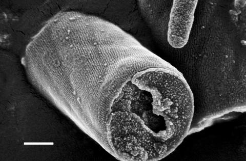
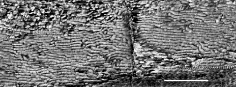
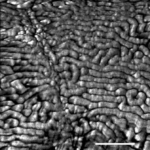
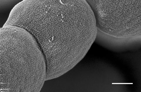
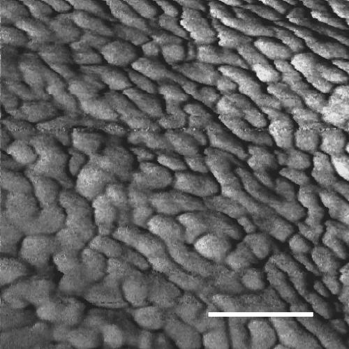
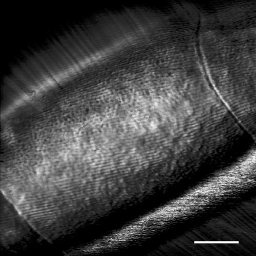
Many filamentous cyanobacteria are motile by gliding, which requires attachment to a surface. There are two main theories to explain the mechanism of gliding. According to the first, the filament is pushed forward by small waves that pass along the cell surface. In the second, gliding is powered by the extrusion of slime through pores surrounding each cell septum. We have previously shown that the cell walls of several motile cyanobacteria possess an array of parallel fibrils between the peptidoglycan and the outer membrane and have speculated that the function of this array may be to generate surface waves to power gliding. Here, we report on a study of the cell surface topography of two morphologically different filamentous cyanobacteria, using field emission gun scanning electron microscopy (FEGSEM) and atomic force microscopy (AFM). FEGSEM and AFM images of Oscillatoria sp. strain A2 confirmed the presence of an array of fibrils, visible as parallel corrugations on the cell surface. These corrugations were also visualized by AFM scanning of fully hydrated filaments under liquid; this has not been achieved before for filamentous bacteria. FEGSEM images of Nostoc punctiforme revealed a highly convoluted, not parallel, fibrillar array. We conclude that an array of parallel fibrils, beneath the outer membrane of Oscillatoria, may function in the generation of thrust in gliding motility. The array of convoluted fibrils in N. punctiforme may have an alternative function, perhaps connected with the increase in outer membrane surface area resulting from the presence of the fibrils.
Figures
References
-
- Adams, D. G. 1997. Cyanobacteria, p. 109-148. In J. A. Shapiro and M. Dworkin (ed.), Bacteria as multicellular organisms. Oxford University Press, Oxford, United Kingdom.
-
- Adams, D. G., and P. Duggan. 1999. Heterocyst and akinete differentiation in cyanobacteria. New Phytol. 144:3-33.
-
- Bhaya, D., N. R. Bianco, D. Bryant, and A. Grossman. 2000. Type IV pilus biogenesis and motility in the cyanobacterium Synechocystis sp. PCC6803. Mol. Microbiol. 37:941-951. - PubMed
Publication types
MeSH terms
LinkOut - more resources
Full Text Sources
Other Literature Sources
Miscellaneous
