. 2007 Aug;19(8):2606-23.
doi: 10.1105/tpc.107.053736. Epub 2007 Aug 10.

A ribonuclease III domain protein functions in group II intron splicing in maize chloroplasts

Affiliations

A ribonuclease III domain protein functions in group II intron splicing in maize chloroplasts

Kenneth P Watkins et al. Plant Cell. 2007 Aug.

Abstract

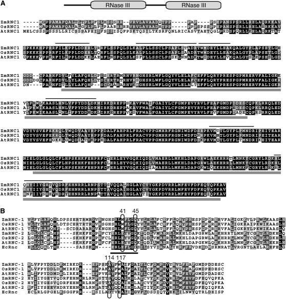
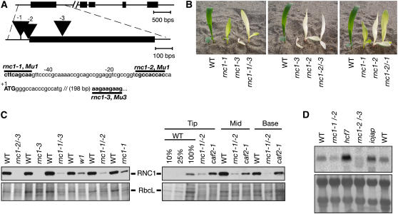
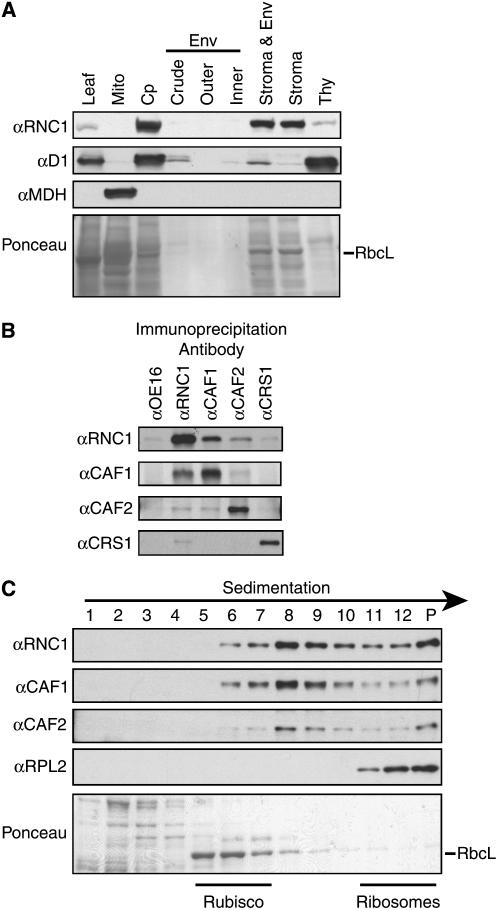
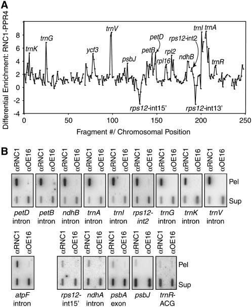
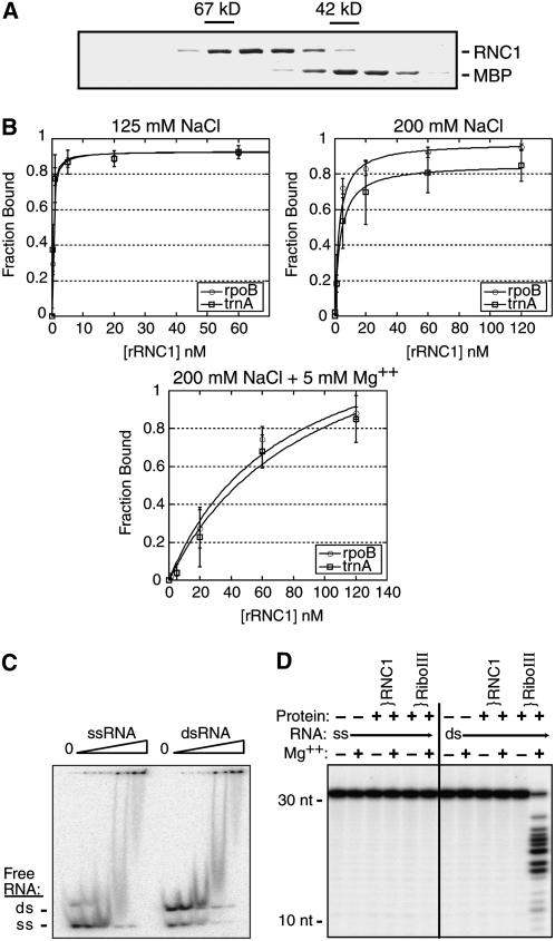
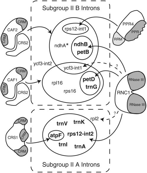
Chloroplast genomes in land plants harbor approximately 20 group II introns. Genetic approaches have identified proteins involved in the splicing of many of these introns, but the proteins identified to date cannot account for the large size of intron ribonucleoprotein complexes and are not sufficient to reconstitute splicing in vitro. Here, we describe an additional protein that promotes chloroplast group II intron splicing in vivo. This protein, RNC1, was identified by mass spectrometry analysis of maize (Zea mays) proteins that coimmunoprecipitate with two previously identified chloroplast splicing factors, CAF1 and CAF2. RNC1 is a plant-specific protein that contains two ribonuclease III (RNase III) domains, the domain that harbors the active site of RNase III and Dicer enzymes. However, several amino acids that are essential for catalysis by RNase III and Dicer are missing from the RNase III domains in RNC1. RNC1 is found in complexes with a subset of chloroplast group II introns that includes but is not limited to CAF1- and CAF2-dependent introns. The splicing of many of the introns with which it associates is disrupted in maize rnc1 insertion mutants, indicating that RNC1 facilitates splicing in vivo. Recombinant RNC1 binds both single-stranded and double-stranded RNA with no discernible sequence specificity and lacks endonuclease activity. These results suggest that RNC1 is recruited to specific introns via protein-protein interactions and that its role in splicing involves RNA binding but not RNA cleavage activity.


Figures

Figure 1.
Figure 1.
The RNC1 Protein. (A) Multiple sequence alignment of maize RNC1 (Zm RNC1) with its rice and Arabidopsis orthologs (Os RNC1 and At RNC1, respectively). The domain organization of RNC1 is shown diagrammatically at top. Identical residues are shaded in black, and similar residues are shaded in gray. Gray bars beneath the alignment mark the two RNase III domains. Lines above the alignment mark the tryptic peptides identified by mass spectrometry in both the CAF1 and CAF2 coimmunoprecipitates; additional peptides were identified in the CAF1 but not the CAF2 immunoprecipitation and vice versa (see Supplemental Table 1 online). At RNC1 corresponds to At4g37510 and Os RNC1 corresponds to Os01g59510. The transit peptide cleavage sites predicted by ChloroP (Emanuelsson and von Heijne, 2001) are after amino acids 28, 30, and 51 for Zm RNC1, Os RNC1, and At RNC1, respectively. (B) Multiple sequence alignment of the two RNase III domains from Zm RNC1, Os RNC1, and At RNC1 with the single RNase III domain from E. coli RNase III. Residues that coordinate a catalytic Mg2+ ion in bacterial RNase III are circled and are numbered according to the E. coli protein. The RNase III signature motif is underlined.
Figure 2.
Figure 2.
Mutant Alleles of rnc1. (A) Positions of Mu transposon insertions in the rnc1 gene. Protein-coding regions are indicated by rectangles, untranslated regions by lines, and insertions by triangles. The name of the mutant allele (rnc1-1, -2, or -3) is shown above each insertion. The positions of the insertions in the wild-type sequence are shown below, with the nine nucleotides that were duplicated during insertion and the name of the specific Mu element marked in boldface. (B) Phenotypes of rnc1 mutant seedlings. The seedlings are homozygous for the rnc1-1, rnc1-2, or rnc1-3 allele or are the heteroallelic progeny of complementation crosses, as indicated. Plants were grown for 9 d. (C) Loss of the RNC1 protein in rnc1 mutant leaf tissue. Immunoblots were probed with the RNC1 antibody (top panels); the same blots stained with Ponceau S are shown below, with the band corresponding to RbcL marked. The left panels show results for the first leaf harvested soon after it emerged from the coleoptile. The right panels show a developmental profile of the second leaf of 7-d-old seedlings; tissue was taken from the leaf base (youngest tissue), middle, and tip (oldest tissue). The albino mutants w1 and caf2 are shown to illustrate the effects on RNC1 protein level that result from the absence of plastid ribosomes. (D) Loss of rnc1 mRNA in rnc1 mutants. An RNA gel blot of total seedling leaf RNA was probed with the rnc1 cDNA. RNA from hcf7 and iojap are shown to illustrate rnc1 RNA levels in mutants with mild (hcf7) or severe (iojap) plastid ribosome deficiencies. An image of the blot stained with methylene blue is shown below.
Figure 3.
Figure 3.
RNC1 Is Associated with CAF1 and CAF2 in the Chloroplast Stroma. (A) Immunoblots of leaf and subcellular fractions. Chloroplasts (Cp) and chloroplast subfractions were from the fractionated chloroplast preparation described and verified previously (Williams and Barkan, 2003); the samples in these lanes are derived from the same initial quantity of chloroplasts. The blot was reprobed to detect a marker for thylakoid membranes (D1) and mitochondria (MDH). Env, envelope; Mito, leaf mitochondria; Thy, thylakoid membranes. (B) Coimmunoprecipitation of RNC1 with CAF1 and CAF2. Stroma was subjected to immunoprecipitation with the antibodies named at top. The presence of specific proteins in the immunoprecipitation pellets was tested by immunoblot analysis with the antibodies listed at left. Immunoprecipitations with OE16 antibody served as a negative control. (C) Cosedimentation of RNC1 with intron ribonucleoprotein particles. Stromal extract was sedimented in sucrose gradients under conditions in which particles of greater than ∼70S pellet (P). An equal volume of each gradient fraction was analyzed by probing immunoblots with the antibody indicated at left. RPL2 is a protein in the large ribosomal subunit and marks the position of ribosomes. The RbcL band in the Ponceau S–stained blot at bottom marks the position of Rubisco. The peaks of CAF1, CAF2, and RNC1 between Rubisco and ribosomes coincide with the positions of several group II intron RNAs (Jenkins and Barkan, 2001; Till et al., 2001).
Figure 4.
Figure 4.
Identification of RNA Ligands of RNC1 by Coimmunoprecipitation Analyses. (A) RIP-chip data displayed to show differential enrichment of RNA sequences in RNC1 versus PPR4 immunoprecipitations. The median log2(F635:F532) for replicate spots across replicate immunoprecipitations with anti-PPR4 antibody (data from Schmitz-Linneweber et al., 2006) was subtracted from the median log2(F635:F532) for replicate immunoprecipitations with RNC1 antibody. The locus name is indicated for peaks that include fragments whose enrichment from wild-type stroma ranked in the top 15th percentile and if the sequences showed significant differential enrichment from either RNC1 versus PPR4 immunoprecipitations or from wild-type versus rnc1-1/rnc1-2 mutant stroma (see Supplemental Table 2 online). (B) Verification of RIP-chip data by slot-blot hybridization. RNA purified from the pellets (Pel) and supernatants (Sup) of RNC1 immunoprecipitation reactions with wild-type stroma was applied to slot blots and hybridized with probes specific for the indicated sequence. Immunoprecipitations with an antibody to OE16 was used as a negative control. The total signal from the pellet plus supernatant is greater for sequences that are strongly enriched in RNC1 immunoprecipitations than in the control immunoprecipitation. This is due in part to the fact that one-sixth of the total pellet sample was analyzed in each slot, whereas only one-twelfth of the corresponding supernatant was analyzed. In addition, the signal is artifactually reduced in the supernatant samples due to saturation of the binding capacity of the membrane with the large amount of RNA.
Figure 5.
Figure 5.
Coimmunoprecipitation of Unspliced tRNA Precursors but Not Spliced tRNAs with RNC1. RNA purified from an RNC1 immunoprecipitation supernatant (Sup) and pellet (Pel) was analyzed on duplicate RNA gel blots by hybridization to the indicated exon-specific and intron-specific probes. Equal fractions of the pellet and supernatant samples were analyzed. An immunoprecipitation with antibody to OE16 served as a negative control. RNA extracted from an equivalent amount of stroma was analyzed for comparison. Blots were initially probed with spliced cDNAs corresponding to the indicated mature tRNAs (exons) and were reprobed with intron-specific probes; residual signal at the positions of mature tRNAs remained after the intron probings. The right panel shows analogous samples probed to detect 16S rRNA. Pre-16S rRNAs were not detectable in any of the lanes.
Figure 6.
Figure 6.
Loss of Plastid Ribosomes and Plastid-Encoded Proteins in rnc1 Mutants. (A) Reduced accumulation of photosynthetic enzyme complexes in rnc1 mutants. Immunoblots of total leaf extract (5 μg of protein or dilutions as indicated) were probed with antibodies to the protein labeled at left. AtpA, D1, PsaD, and PetD are subunits of ATP synthase, photosystem II, photosystem I, and the cytochrome b6f complex, respectively. The same blot stained with Ponceau S is shown below to illustrate the relative loading of the samples and the abundance of RbcL. (B) Loss of plastid rRNAs in rnc1 mutants. Total seedling leaf RNA (0.5 μg) from the genotype indicated at top was analyzed by RNA gel blot hybridization using probes specific for the RNAs indicated at bottom. The pigmentation of the mutants is indicated at top: iv, ivory leaves; pg, pale green leaves. One of the blots stained with methylene blue is shown below to illustrate equal loading of the cytosolic rRNAs.
Figure 7.
Figure 7.
Chloroplast Splicing Defects in rnc1 Mutants. (A) Poisoned primer extension assays for mRNA splicing. Reverse transcriptase reactions were initiated with radiolabeled primers complementary to the exon downstream of the indicated intron; a dideoxy nucleotide that terminates reverse transcription after different distances on spliced and unspliced templates was included in the reactions. The splicing defects for the petB, petD, and ndhB introns were confirmed with RNase protection assays (see Supplemental Figure 2 online). The asterisk at the petB panel marks a product that terminates near the branch point adenosine; this likely originates from the lariat intermediate that is the product of the first step in splicing. Data are shown for the weak allele combination rnc1-1/rnc1-2 to minimize secondary effects due to the loss of plastid ribosomes. P, primer; S, spliced; U, unspliced. (B) RNA gel blot analysis of tRNA splicing. Blots were probed with cDNAs derived from the indicated spliced tRNA. Asterisks mark the positions of unspliced precursors, as deduced from their size and by their hybridization to intron-specific probes (Figure 5; data not shown). The trnR-ACG gene does not contain an intron; it is shown because it emerged as a possible RNC1 ligand in the RIP-chip assays and to illustrate that tRNA metabolism is not globally disrupted in rnc1 mutants. iv, ivory leaves; pg, pale green leaves. (C) RNase protection assays of mRNA splicing. Probes spanned either the 3′ splice junction (rps16, rpl16, and ycf3-int2) or the 5′ splice junction (ndhA and ycf3-int1). The asterisk marks a product anticipated to result from hybridization to the excised intron.
Figure 8.
Figure 8.
RNA Gel Blots Showing the Levels of Excised Introns in rnc1 Mutants. Total leaf RNA (5 μg per lane) was analyzed by RNA gel blot hybridization using probes specific for the indicated intron. RNA from a caf1 mutant, which has a strong defect in petD splicing, was included on the petD intron blot for comparison. Arrows point to presumed excised introns: their sizes match those of excised introns and they do not hybridize to exon probes (data not shown). The reduced splicing of the rpl2 and rps12-int2 introns in the strong rnc1 alleles (rnc1-2/rnc1-3 and rnc1-2 homozygotes) is not informative because these introns are in subgroup IIA and fail to splice in all maize mutants lacking plastid ribosomes. iv, ivory leaves; pg, pale green leaves.
Figure 9.
Figure 9.
In Vitro Assays with Recombinant RNC1. (A) Elution of untagged rRNC1 from the gel filtration column. Fractions from the gel filtration column were analyzed by SDS-PAGE, and proteins were detected with Coomassie blue. Contiguous fractions from the relevant portion of the elution are shown. The elution of rRNC1 compared with BSA (67 kD) and MBP (42 kD) indicates that it is monomeric (predicted molecular mass, 59 kD). The peak rRNC1 fractions were pooled, and residual MBP was removed by passage over an amylose affinity column prior to use in in vitro assays. (B) Filter binding assays showing RNA binding activity of rRNC1. Assays were performed with trace amounts of radiolabeled RNAs corresponding to an RNC1 ligand (trnA intron) and an RNA of similar size that did not show evidence of interacting with RNC1 in vivo (rpoB-coding region). RNA was heated in the absence of salts and then snap-cooled in the presence of 125 mM NaCl (top panel), slow-cooled in 200 mM NaCl (middle panel), or slow-cooled in 200 mM NaCl and 5 mM Mg2+ (bottom panel). Single-site binding isotherms were fit to the data using the following equation: fraction RNA bound = (maximum RNA bound × [protein])/(Kd + [protein]). Data points are averages from three assays, with error bars indicating 1 sd. (C) Gel mobility shift assay showing the relative affinity of rRNC1 for single-stranded and double-stranded RNA. A synthetic RNA oligonucleotide was radiolabeled and either heated and snap-cooled (ssRNA) or mixed with a twofold excess of its complement and slow-cooled in the presence of monovalent salts to promote duplex formation (dsRNA). The annealing was not complete, so the dsRNA substrate includes both dsRNA and ssRNA. RNA (40 pM) was incubated with increasing concentrations of rRNC1 (32, 80, 200, and 500 nM). The disappearance of unbound RNA is used to monitor protein binding. (D) rRNC1 lacks RNase III–like endonuclease activity. RNA substrates were the same synthetic ssRNA and dsRNA 31-mers used in (C). Reactions contained rRNC1 at a concentration of 125 nM or E. coli RNase III (RiboIII) at 10 nM (dimer concentration).
Figure 10.
Figure 10.
Nucleus-Encoded Chloroplast Splicing Factors in Maize and Their Intron Targets. The 17 group II introns in the maize chloroplast genome are divided into subgroups IIA and IIB according to Michel et al. (1989). Nucleus-encoded splicing factors are shown, with identified domains shaded. The designated intron targets (solid arrows) have been shown to coimmunoprecipitate with the indicated proteins and to require those proteins for optimal splicing in vivo. Dashed arrows designate interactions detected by coimmunoprecipitation but for which a role in splicing has not been documented. Results are summarized from this work and from Jenkins et al. (1997), Vogel et al. (1999), Till et al. (2001), Ostheimer et al. (2003), and Schmitz-Linneweber et al. (2005, 2006). The Arabidopsis orthologs of CAF1, CAF2, and CRS1 have analogous functions (Asakura and Barkan, 2006). Arabidopsis HCF152 is the only other putative chloroplast splicing factor reported in land plants (Meierhoff et al., 2003); HCF152 is required for the accumulation of spliced petB RNA, but not for the accumulation of excised petB intron, so its role in splicing is not firmly established. In addition to these nucleus-encoded splicing factors, a role for plastid-encoded MatK in subgroup IIA splicing has been proposed (reviewed in Bonen and Vogel, 2001). * The ndhA intron shows a strong interaction with and dependence on CAF2 and a weak interaction with and dependence on CAF1.

Similar articles

Cited by

References

    1. Asakura, Y., and Barkan, A. (2006). Arabidopsis orthologs of maize chloroplast splicing factors promote splicing of orthologous and species-specific group II introns. Plant Physiol. 142 1656–1663. - PMC - PubMed
    1. Barkan, A. (1993). Nuclear mutants of maize with defects in chloroplast polysome assembly have altered RNA metabolism. Plant Cell 5 389–402. - PMC - PubMed
    1. Barkan, A. (1998). Approaches to investigating nuclear genes that function in chloroplast biogenesis in land plants. Methods Enzymol. 297 38–57.
    1. Barkan, A., Klipcan, L., Ostersetzer, O., Kawamura, T., Asakura, Y., and Watkins, K. (2007). The CRM domain: An RNA binding module derived from an ancient ribosome-associated protein. RNA 13 55–64. - PMC - PubMed
    1. Barkan, A., Walker, M., Nolasco, M., and Johnson, D. (1994). A nuclear mutation in maize blocks the processing and translation of several chloroplast mRNAs and provides evidence for the differential translation of alternative mRNA forms. EMBO J. 13 3170–3181. - PMC - PubMed

Publication types

MeSH terms

Associated data

LinkOut - more resources